On this page
Quick Reference
Overview and Recommendations
Background
- • (PG) is a rare neutrophilic dermatosis causing rapidly progressive, intensely painful, sterile cutaneous ulceration. Classic lesions have a necrotic base and reddish-violaceous, undermined borders; minor trauma may enlarge them through .
- •PG is noninfectious, although open ulcers can develop colonization or secondary infection. A positive wound culture does not by itself exclude PG; in one cohort, bacterial growth occurred in 50.3% of PG ulcers and the organisms did not distinguish them from venous ulcers.
- •Recognize PG as a , not a diagnosis established by a pathognomonic biopsy. Histology may show dermal neutrophilic inflammation, necrosis, and ulceration, but findings are variable and nonspecific; biopsy chiefly helps exclude infection, , malignancy, and other ulcerative disorders.
- •Classic ulcerative PG is the prototypic phenotype, but bullous, pustular, vegetative, peristomal, postoperative, and genital forms occur. Phenotypes may overlap or evolve, so classify the morphology while correlating it with lesion tempo, comorbidity, microbiology, and histology when needed.
- •PG commonly occurs without an identifiable systemic disorder, but approximately 50%-70% of patients in some series have an associated condition. Ask about , inflammatory arthritis, hematologic disease, and childhood-onset autoinflammatory syndromes; an initially negative history does not end longitudinal surveillance.
Evaluation
- •Assess the lesion’s trajectory from its initial tender papule, pustule, or nodule through rapid ulceration. Severe pain, rapid progression, necrosis, an irregular reddish-violaceous or gunmetal border, and undermining strongly support PG, but none is pathognomonic.
- •Examine the entire lesion and surrounding skin for new ulcers, active undermining, erythema, drainage, exposed tendon or muscle, and pathergy at sites of biopsy, venipuncture, surgery, injections, appliance changes, or other trauma. Document distribution; lower legs are most common, but PG can occur at any cutaneous site.
- •Treat fever, malaise, leukocytosis, and elevated inflammatory markers as nonspecific. Assess the clinical trajectory, because antibiotics and source-control procedures may fail to halt sterile inflammation while repeated trauma can accelerate it.
- •For postoperative or peristomal lesions, evaluate urgently for infection while considering PG. Wound separation, necrosis, purulent discharge, severe pain, extension beyond an incision or stoma, or worsening after debridement should prompt dermatology and surgical reassessment.
- •Obtain a with differential and review the peripheral smear. Measure and/or , electrolytes, glucose, albumin, liver enzymes, bilirubin, and renal function to establish inflammatory and treatment-risk baselines.
- •Ask about diarrhea, rectal bleeding, abdominal pain, weight loss, perianal disease, and prior bowel surgery. Ask about inflammatory back pain, synovitis, prolonged morning stiffness, enthesitis, dactylitis, and episodic arthritis; arrange gastroenterology or rheumatology assessment when findings support associated disease.
- •Consider hematologic assessment for bullous or atypical PG, unexplained cytopenias, abnormal smear findings, constitutional symptoms, recurrent disease, or known myeloid disease. Review for myelodysplastic syndrome, leukemia, lymphoma, myeloproliferative disease, and when the clinical context warrants it.
- •Order targeted studies for when lesions are palpable or retiform purpura, acral or multifocal, or accompanied by renal, pulmonary, neurologic, ocular, or constitutional findings. Use vascular studies, including ankle-brachial index or toe pressures and arterial or venous , when ischemia or venous disease is plausible.
- •Use an incisional or punch through the active ulcer edge into adjacent normal-appearing skin when morphology is atypical, infection is plausible, or vasculitis or malignancy cannot be excluded. Add base tissue and coordinate routine histology with bacterial, fungal, and mycobacterial studies when deep infection, an infiltrative process, or malignancy is suspected; explain pathergy risk before sampling.
- •Interpret histology as primarily exclusionary. A nondiagnostic biopsy does not exclude PG, while organisms, vasculitis, atypical cells, or an unexpected infiltrate should redirect the workup before immunosuppression.
- •Use deep tissue or properly collected wound specimens when infection is clinically suspected rather than relying on a superficial swab. Culture positivity may indicate colonization or secondary infection; treat infection when clinical, microbiologic, histologic, or imaging findings support invasion.
- •Apply structured diagnostic reasoning rather than relying on one finding. The 2018 Delphi framework requires one major criterion plus at least four minor criteria, while emphasizes progressive disease, a reddish-violaceous border, and reasonable exclusion of other diagnoses; a score of 10 or more has traditionally supported PG, but does not eliminate infection, vascular disease, malignancy, or mixed pathology.
Management
- •Use local treatment only when disease is limited, superficial, nonprogressive, and located where atraumatic care will not jeopardize function. Reassess frequently and escalate when new ulcers, advancing undermining, substantial depth, severe functional impairment, extracutaneous disease, or systemic inflammation develops.
- •Cleanse gently with lukewarm saline or water without rubbing the active border. Apply a nonadherent contact layer, such as petrolatum-impregnated gauze or a soft silicone interface, with an absorbent secondary dressing; maintain a moist rather than macerated or desiccated environment.
- •Avoid hydrogen peroxide, hypochlorite, iodine, and other caustic antiseptics on the active ulcer unless a wound specialist has a specific, time-limited indication. Secure dressings without adhesive across the ulcer edge, remove them slowly after moistening, and avoid repeated manipulation.
- •For limited superficial inflammation, a potent topical corticosteroid such as may be applied to the active edge and surrounding skin under dermatology direction. is a steroid-sparing option for thin skin, near an ostomy, or when corticosteroid atrophy is a concern; evidence does not establish an optimal dose, frequency, or monotherapy regimen.
- •Treat pain before dressing changes. Use an ulcer-compatible topical anesthetic according to product labeling and add systemic analgesia when needed; record pain on a reproducible 0-10 scale and address sleep, mobility, nutrition, mood, and dressing tolerance.
- •Control edema and venous hypertension only after confirming adequate arterial supply and at a tolerable compression level. Elevate the limb, mobilize when feasible, protect the periwound skin, and manage peristomal leakage, friction, and appliance trauma with an experienced ostomy and wound-care team.
- •Start systemic therapy for rapidly enlarging, painful, deep, multifocal, function-threatening, extracutaneous, or substantially inflammatory disease. Reassess progression, pain, ulcer area, depth, and new lesions at least weekly during induction; the first goals are cessation of enlargement and new lesions, followed by reduced pain and undermining and then epithelial advancement.
- •For severe active disease, use 20-40 mg orally once daily, approximately 0.6-0.8 mg/kg/day, maintaining the starting dose until enlargement and inflammatory edge activity have clearly stopped, usually for 2-4 weeks, then taper slowly according to response. Avoid prolonged high-dose corticosteroid monotherapy and monitor glucose, blood pressure, infection, mood, myopathy, bone risk, and adrenal suppression.
- •Use 3-5 mg/kg/day orally in two divided doses when rapid steroid-sparing control is needed, particularly with diabetes, severe steroid toxicity, or a corticosteroid contraindication. Check creatinine/eGFR, urea, potassium, magnesium, liver tests, blood pressure, and drug interactions at baseline and at least every 1-2 weeks during dose adjustment, then monthly when stable; avoid it in uncontrolled infection, significant renal impairment, uncontrolled hypertension, or untreated malignancy risk.
- •For fulminant disease, unreliable oral absorption, or a threatened critical site, intravenous methylprednisolone commonly 500-1000 mg intravenously once daily for 3 consecutive days can be used, although PG-specific comparative evidence for pulse therapy is weak. Choose prednisone or cyclosporine by comorbidity and toxicity rather than assuming a difference in efficacy.
- •Add a steroid-sparing agent before repeated high-dose corticosteroid courses when disease relapses during tapering. Options include 500 mg orally twice daily, increasing over 1-2 weeks to 1-1.5 g twice daily if tolerated; 15-25 mg orally or subcutaneously once weekly with folic acid; or 1-2.5 mg/kg orally once daily, with monitoring and reproductive-risk review.
- •Consider when rapid biologic treatment is needed, particularly with inflammatory bowel disease or inflammatory arthritis. Use 5 mg/kg intravenously at weeks 0, 2, and 6, then every 8 weeks when ongoing suppression is required; screen for tuberculosis and hepatitis B, review infection risk, and assess for heart failure and demyelinating disease.
- •Use when outpatient self-administration or treatment of inflammatory bowel disease or arthritis favors a subcutaneous agent: 160 mg subcutaneously at week 0, 80 mg at week 2, then 40 mg every 2 weeks. Before anti-TNF therapy, perform symptom-directed infection assessment, tuberculosis evaluation, hepatitis B testing, CBC, liver tests, and vaccination review; do not start during uncontrolled serious infection and avoid live vaccines during therapy.
- •Reserve intravenous immunoglobulin, ustekinumab, anakinra, canakinumab, or IL-23 inhibition for refractory disease or selected comorbid and autoinflammatory contexts. Examples include intravenous immunoglobulin 2 g/kg divided over 2-5 days, approximately 6 mg/kg intravenously once followed by 90 mg subcutaneously at week 8 and every 8-12 weeks, 100 mg subcutaneously once daily, or 150 mg subcutaneously every 8 weeks; evidence is mainly observational or case-based.
- •Do not present IL-17 or IL-23 inhibition as established first-line PG treatment. IL-17 blockade can aggravate or destabilize inflammatory bowel disease; selective IL-23 inhibition may be considered after conventional-treatment failure, such as 100 mg subcutaneously at weeks 0 and 4 then every 8 weeks, with explicit recognition that treatment is off-label for PG.
- •Avoid routine aggressive debridement, wide excision, and primary closure while the border is active or the diagnosis remains uncertain. Use gentle cleansing and selective removal of loose necrotic material; operate only for demonstrable source control, deep or prosthetic infection, sepsis, compartment or limb threat, uncontrolled hemorrhage, or threatened function, removing no more tissue than necessary.
- •Defer , , biologic matrix, and reconstructive revision until inflammation is controlled, the wound is stable and adequately perfused, and a perioperative immunosuppressive plan is in place. Minimize incision, shear, adhesive, donor-site, and dressing trauma, and monitor incision, drain, graft, and venipuncture sites for pathergy.
- •Continue immunosuppression until control is durable: no enlargement or new lesions, settled edge inflammation, sustained pain improvement, and progressive epithelial advancement. Taper deliberately rather than abruptly, record wound area, depth, photographs, pain, function, treatment toxicity, and associated-organ disease, and educate patients to report renewed pain, pustulation, violaceous undermining, or a new ulcer promptly.
Deep Dive — Evidence Details
Pyoderma Gangrenosum: Definition, Nosology, and Clinical Identity
- ▸Rapidly progressive, intensely painful ulceration with a necrotic base and reddish-violaceous undermined border should raise concern for classic PG, and pathergy means minor trauma or reflexive debridement may worsen it.
- ▸A positive wound culture does not by itself exclude PG because established ulcers may acquire secondary microbial growth; interpret cultures as evidence of possible superinfection rather than proof of an infectious origin.
- ▸PG is a clinicopathologic diagnosis: biopsy findings are variable and nonspecific, so use biopsy primarily to help exclude infection, cutaneous vasculitis, malignancy, or another ulcerative disorder, while balancing the risk of pathergy.
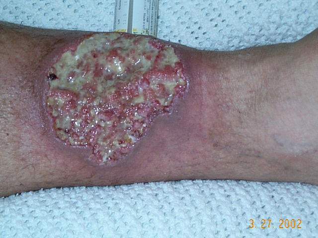

(PG) is a rare neutrophilic dermatosis defined clinically by rapidly progressive, intensely painful, sterile cutaneous ulceration, usually with a necrotic base and a reddish-violaceous, undermined border. The ulcer may enlarge after minor trauma, a phenomenon called , because tissue injury can amplify the underlying neutrophil-dominant inflammation rather than initiate infection. [2][5][7]
The name is historically misleading. “Pyoderma” suggests pus-producing infection, and “gangrenosum” suggests primary tissue necrosis from an infectious or ischemic process; neither describes the usual mechanism. PG is a noninfectious inflammatory disease, although an established ulcer can acquire secondary microbial growth, and a positive wound culture therefore does not by itself exclude PG. [4][7] Treat cultures as evidence of possible superinfection, not as proof that the original ulcer is infectious.
PG is a , not a diagnosis made by a pathognomonic biopsy. Histology may show dermal neutrophilic inflammation, necrosis, and ulceration, but these findings are variable and nonspecific; chronic ulcers may lack the acute neutrophilic pattern, and neutrophils can also occur in ordinary chronic wounds. Biopsy is most useful when it helps exclude infection, , malignancy, or another ulcerative disorder, particularly when the clinical pattern is atypical. Balance that diagnostic value against pathergy when choosing the biopsy site and technique. [2]
PG belongs to the broader spectrum, in which sterile neutrophilic inflammation can produce clinically different lesions rather than one uniform morphology. The spectrum includes , acute febrile neutrophilic dermatosis with papules, plaques, or nodules, alongside PG and more localized or pustular-neutrophilic phenotypes such as and . This relationship is mechanistic and clinicopathologic: related inflammatory biology may appear as superficial plaques, pustules, sterile abscess-like collections, or destructive ulceration, so the presence of neutrophils alone does not establish PG. [1][2]
The term PG has practical limits. Classic ulcerative PG is the dominant and best-recognized phenotype, but clinicians also use the labels atypical or bullous, pustular, and vegetative PG for lesions whose depth, surface, tempo, or clinical context differs from the classic ulcer. These labels describe clinical variants within the PG spectrum rather than independent infectious entities; variant assignment should follow careful correlation of morphology, evolution, comorbidity, microbiology, and, when needed, histology. [5][19]
| Term | Defining clinical concept | Relationship to PG | Major diagnostic implication |
|---|---|---|---|
| Classic ulcerative PG | Rapidly progressive, painful ulceration with a necrotic base and undermined violaceous border; pathergy may occur. [2][5] | The prototypic PG phenotype. [19] | Prioritize PG while actively excluding infection, vascular disease, vasculitis, and malignancy; avoid reflexive debridement because trauma may worsen the ulcer. [2][7] |
| Atypical or bullous PG | Superficial hemorrhagic bullae or bullous ulceration, often associated with hematologic disease. [5][12] | A recognized PG phenotype with a different surface morphology from classic ulcerative disease. [5][12] | Consider a neutrophilic process in a patient with leukemia or another hematologic disorder; culture and biopsy may be required to exclude infection or other bullous disorders. [2][12] |
| Pustular PG | Pustular lesions, reported particularly during inflammatory bowel disease flares. [5] | A pustular expression of the PG spectrum rather than the usual deep ulcerative form. [5] | Do not equate pustular drainage with bacterial pyoderma; interpret it alongside disease tempo, pain, pathergy, and systemic context. [4][5] |
| Vegetative PG | A more indolent, often solitary ulcerative lesion with less prominent violaceous change and relatively limited systemic inflammation. [5] | A chronic, less destructive PG phenotype. [5] | The muted appearance can lower clinical suspicion; maintain a broad differential and use biopsy when malignancy or another chronic ulcer cause remains plausible. [2][5] |
| Sweet syndrome | Acute febrile neutrophilic dermatosis presenting with inflammatory papules, plaques, or nodules rather than a primary destructive ulcer. [1] | A related neutrophilic dermatosis, not synonymous with PG. [1] | Fever and neutrophilic lesions without the characteristic progressive ulcer and undermined border should prompt consideration of Sweet syndrome rather than automatic labeling as PG. [1][2] |
| Neutrophilic dermatosis of the dorsal hands | Localized neutrophilic inflammation involving the dorsal hands, which may be pustular, pseudovesicular, or ulcerative. [1] | A localized phenotype within the neutrophilic dermatosis spectrum that may clinically overlap with PG. [1] | Site and morphology matter; a hand lesion should not be called PG solely because biopsy shows neutrophils. [1][2] |
| Aseptic abscess syndrome | Sterile, neutrophil-rich abscess-like inflammation that can mimic bacterial abscesses. [1] | A related sterile neutrophilic inflammatory disorder, but not interchangeable with cutaneous PG. [1] | Failure to improve with appropriate antibiotics and repeatedly negative cultures should reopen the diagnosis and prompt assessment for a neutrophilic disorder. [1][4] |
Thus, PG is best recognized as a clinical pattern of sterile, neutrophil-mediated ulceration whose diagnosis depends on the whole trajectory of the lesion and the disciplined exclusion of mimics, not on the word “pyoderma,” a positive culture from a chronic wound, or neutrophilic inflammation in isolation. [2][4]
Innate Immune Dysregulation and Pathogenesis of Pyoderma Gangrenosum
- ▸PG is a self-amplifying myeloid-neutrophil inflammatory response in which IL-1, IL-8, and TNF recruit and activate neutrophils, whose tissue injury generates further danger signals.
- ▸Pathergy means trauma can initiate or enlarge PG lesions, so repeated surgical debridement may produce progressive wound expansion rather than source control.
- ▸A positive wound culture does not establish microbial causation: bacterial growth occurred in 50.3% of PG ulcers, the same proportion observed in venous ulcers, and culture positivity alone should not justify repeated destructive procedures.
is best understood as a disorder in which tissue injury is converted into an excessive, self-amplifying , rather than as a primary microbial disease. The central cellular abnormality is functional dysregulation of the myeloid-neutrophil axis: activated monocytes and macrophages produce inflammatory mediators, the skin recruits and activates neutrophils, and the resulting tissue injury releases additional endogenous danger signals that sustain inflammation. Available evidence supports this model, but does not establish one uniform molecular pathway for every PG phenotype; inter-individual variability and the rarity of the disease make much of the mechanistic literature observational, transcriptomic, or derived from genetically defined syndromes [21][22].
A useful sequence begins with innate immune sensing. Tissue damage or an inflammatory trigger can activate -dependent transcription in myeloid cells, increasing expression of proinflammatory genes such as IL1B and TNF. A second signal can assemble an , an intracellular protein complex that converts precursor interleukin-1β into its active form and can promote inflammatory cell death. Interleukin-1β then acts on keratinocytes, endothelial cells, fibroblasts, and leukocytes, inducing further inflammatory gene expression and chemokine production. This provides a biologic explanation for the clinical behavior of PG: a small initiating insult can generate a disproportionately intense local response, while tissue destruction supplies new danger signals that perpetuate the cycle. IL-1 is linked experimentally and clinically to inflammasome activation, keratinocyte signaling, and neutrophil recruitment in neutrophil-rich dermatoses [23].
are therefore effectors, not merely histologic bystanders. Interleukin-8 (CXCL8) establishes a chemotactic gradient that draws neutrophils into lesional skin; activated neutrophils then release proteases, reactive oxygen species, and extracellular chromatin structures that damage surrounding tissue and impair orderly repair. The available human data connect this model to PG: IL8 is elevated in PG lesional skin, and in patients with the PG-causing OTULIN-R57C variant, IL8 and IL1B were selectively upregulated in monocytes and were accompanied by increased plasma concentrations [21]. The consequence is a feed-forward loop in which myeloid cytokine production recruits neutrophils, neutrophil injury enlarges the tissue danger field, and the injured tissue reactivates myeloid cells.
Tumor necrosis factor (TNF) can reinforce this loop at several levels by activating inflammatory transcription, promoting endothelial and leukocyte activation, and increasing susceptibility of resident cells to inflammatory death. TNF is elevated in blood and lesions in some patients with PG, although this finding is not universal and should not be interpreted as proof of a single TNF-driven disease subtype [21]. The OTULIN-R57C kindreds provide unusually direct human mechanistic evidence: the mutation preserved OTULIN’s catalytic activity but disrupted its binding to the linear ubiquitin assembly complex, while patient monocytes showed heightened IL1B expression, impaired suppression of inflammasome activity, elevated TNF, and dermal fibroblast susceptibility to TNF-dependent cell death [21]. This observation links a defect in ubiquitin-dependent inflammatory regulation to both myeloid cytokine excess and vulnerability of skin-resident cells, offering a coherent explanation for ulceration without requiring invasive infection.
The / axis may represent a second inflammatory circuit that connects innate activation to tissue persistence. IL-23 supports expansion and maintenance of IL-17-producing lymphocytes, whereas IL-17 acts on keratinocytes and stromal cells to induce neutrophil-recruiting mediators. In PG, evidence for this axis is less mature than the evidence for neutrophil recruitment and IL-1 signaling. A 2026 integrative multi-omics study identified shared IL-17, TNF, and cytokine-signaling modules in PG and inflammatory bowel disease, but its conclusions were generated by computational integration rather than direct pathway perturbation [22]. Similarly, a multicenter retrospective study of selective IL-23 inhibition reported clinical improvement in refractory PG, but treatment response cannot by itself prove that IL-23 is the initiating lesion-level abnormality [25]. The most defensible interpretation is that IL-23/IL-17 signaling may maintain or amplify selected PG inflammatory states, potentially interacting with the IL-1, IL-8, and TNF networks rather than replacing them.
Genetic susceptibility is real but heterogeneous. Familial clustering and rare monogenic disorders show that abnormal regulation of signaling, myeloid inflammatory responses, or linear ubiquitin biology can produce PG, but these variants account for only a small proportion of unselected cases [21]. Gain-of-function PSTPIP1 variants occur in PAPA (pyogenic arthritis, PG, and acne), PASH (PG, acne, and suppurative hidradenitis), and related PSTPIP1-associated inflammatory syndromes because PSTPIP1 regulates pyrin inflammasome activity; MEFV variants that impair pyrin inhibition can cause PAAND (pyrin-associated autoinflammation with neutrophilic dermatosis) [21]. The OTULIN-R57C report extends this spectrum by showing that a defect in protein-complex recruitment, rather than loss of catalytic activity, can cause isolated childhood-onset PG [21]. These syndromes demonstrate biological plausibility, not a basis for assigning a monogenic cause to most adult PG.
The same framework explains why PG clusters with inflammatory bowel disease, inflammatory arthritis, and hematologic disease without implying that any one comorbidity is required. These conditions can converge on barrier injury, myeloid activation, cytokine excess, or altered neutrophil handling; alternatively, they may share susceptibility alleles or an inflammatory environment that lowers the threshold for cutaneous disease. A systematic review found inflammatory bowel disease, hematologic disorders, and arthritis among the most frequent systemic associations, but emphasized that observational studies establish association more reliably than causality [19]. Hematologic disease may be especially relevant to bullous PG and myeloid dysfunction, whereas bowel and joint inflammation may provide distinct systemic cytokine contexts; these clinical patterns should be treated as clues to shared immunobiology, not as mechanistic subtypes established in every patient [19][21].
is the clinical expression of this lowered inflammatory threshold: trauma initiates a new lesion or intensifies an existing one. The initiating injury exposes danger-associated molecular signals, disrupts the barrier, activates local keratinocytes and stromal cells, and recruits neutrophils; in susceptible skin, the response fails to contract and resolve. Dysregulated wound healing then becomes part of the disease rather than a neutral repair process, because inflammatory cell death and extracellular-matrix injury create further signals for recruitment. Reports of PG in PAPA syndrome specifically describe pathergy together with impaired wound healing, supporting this biologically plausible connection, although the mechanistic evidence remains largely clinical and syndromic [20]. Surgical trauma can therefore enlarge disease through the same process, which explains why repeated debridement may produce progressive wound expansion rather than source control [7][17].
A positive culture does not invalidate this model. An open ulcer loses its barrier and can acquire organisms from the surrounding skin, environment, or wound care; microbial growth may coexist with sterile neutrophilic inflammation and may represent colonization or secondary infection rather than the cause of lesion initiation. In a retrospective cohort, bacterial growth occurred in 50.3% of PG ulcers, the same proportion observed in venous ulcers, and the organisms isolated did not distinguish the two ulcer types [4]. Interpret cultures in relation to systemic infection, tissue invasion, bacteremia, and the clinical trajectory; do not use culture positivity alone to justify repeated destructive procedures. The inflammatory lesion can remain active after antimicrobial therapy because the principal driver is dysregulated host inflammation, while true secondary infection may still require separate treatment [4].
Pearl: PG behaves as a self-amplifying wound-inflammation circuit: myeloid activation and inflammasome signaling generate IL-1, IL-8, and TNF; these recruit and activate neutrophils, with IL-23/IL-17 signaling potentially sustaining selected disease states; trauma and debridement can feed the circuit, and microbial growth may be a passenger rather than the driver.
Who Develops Pyoderma Gangrenosum: Epidemiology, Associations, and Risk Context
- ▸PG onset is concentrated between 30 and 55 years, with women affected slightly more often than men.
- ▸Approximately 50%-70% of patients in some series have an associated systemic disorder, but a negative systemic history at presentation does not lower the diagnostic probability of PG or eliminate the need for longitudinal review.
- ▸IBD, rheumatoid arthritis, seronegative spondyloarthritis, and hematologic disease are the strongest recurring associations; reported frequencies such as IBD 21.8%, rheumatoid arthritis 15.6%, and seronegative spondyloarthritis 9.4% should guide suspicion rather than be treated as universal prevalence estimates.
(PG) is rare, with an estimated incidence of approximately 3-10 cases per million people per year. It can occur at any age, but onset is concentrated in adulthood, particularly between 30 and 55 years; women are affected slightly more often than men. [5][33] The apparent increase in diagnoses in some health systems should be interpreted cautiously: multicenter Italian data suggest that improved recognition and wider use of structured diagnostic frameworks, rather than a proven rise in true incidence, may explain much of the trend. [34]
A useful clinical distinction is between sporadic PG, in which no systemic disorder is identifiable, and disease-associated PG, in which PG occurs alongside an inflammatory, hematologic, neoplastic, or autoinflammatory condition. Reported cohorts differ because referral patterns and definitions differ, but approximately 50%-70% of patients in some series have an associated systemic disorder; therefore, many patients have no identifiable association. [5] The association is temporally variable. PG may precede the systemic diagnosis, appear concurrently, or develop afterward; a reported patient developed PG five months before myelodysplastic syndrome was recognized. [17] A negative systemic history at presentation therefore lowers neither the diagnostic probability of PG nor the need to review the history longitudinally.
The strongest recurring associations are with (IBD), inflammatory arthritis, and hematologic disease. In a recent Spanish case series, IBD occurred in 21.8% of patients, rheumatoid arthritis in 15.6%, and seronegative spondyloarthritis in 9.4%; a broader systematic review likewise identified IBD, hematologic disorders, and arthritis as the most frequent categories. [19][33] These figures describe selected observational populations rather than the risk conferred by each disease, so they should guide clinical suspicion rather than be treated as universal prevalence estimates.
PG associated with IBD may accompany either or and may occur during active intestinal disease or independently of bowel symptoms. Peristomal lesions are a recognized context, but a stoma or prior abdominal operation does not establish causation. In a multicenter cohort of patients with IBD and PG, 50% had undergone prior surgery and 68% had a stoma in their medical history; these findings identify a population in which postoperative and peristomal disease deserves attention, not a reason to avoid necessary surgery. [35]
The rheumatologic spectrum includes and seronegative inflammatory disease, particularly and IBD-associated spondylitis. Arthritis may be clinically obvious, but a history of inflammatory back pain, enthesitis, recurrent peripheral synovitis, or established IBD can provide the clue when the joint diagnosis is not yet formalized. The association is syndromic rather than diagnostic: most patients with inflammatory arthritis do not develop PG, and PG alone does not identify a particular rheumatologic disease. [19][33]
Hematologic associations include myeloid malignancies, , leukemia, lymphoma, and myeloproliferative disorders. They are less frequent than IBD in most series but clinically consequential because bullous or atypical PG has a recognized relationship with myeloproliferative disease and because PG can precede detection of a hematologic disorder. [5][17][19] is an additional, uncommon association reported in PG cohorts; its presence should be treated as a contextual clue rather than proof that the skin disease is paraneoplastic. [33]
Rare inherited autoinflammatory syndromes are especially relevant when PG begins in childhood or adolescence, recurs with sterile inflammation, or coexists with characteristic extracutaneous disease. PAPA syndrome, pyogenic arthritis, PG, and acne, is caused by pathogenic variants in and typically presents with recurrent sterile arthritis, severe acne, and PG from childhood or adolescence. [27] PASH-spectrum disorders, PG, acne, and hidradenitis suppurativa, with related combinations such as PAPASH when pyogenic arthritis is present, belong to the same rare clinical spectrum. These syndromes account for a small minority of PG and should not be used to explain ordinary adult-onset disease without the relevant phenotype. [33]
Medication exposure and recent tissue injury are risk contexts, not diagnoses. PG has been reported after initiation of biologic therapies used for rheumatic disease, and pharmacovigilance data have generated disproportionate-reporting signals for several biologic classes; spontaneous-reporting analyses cannot establish incidence, causality, or a class-wide contraindication. [38] Recent surgery, a stoma, injections, venipuncture, and other trauma may precede lesions through pathergy, but the temporal relationship does not distinguish PG from infection on its own. Evaluate the lesion trajectory and morphology rather than attributing postoperative ulceration automatically to wound infection or to the medication exposure. [17][35]
| Associated disorder | Approximate strength or frequency of association | Clinical clues | Recommended relevance to evaluation |
|---|---|---|---|
| Commonest systemic category; approximately 20%-30% in reported PG series | Crohn disease or ulcerative colitis; abdominal symptoms; peristomal or pustular PG | Ask about bowel symptoms, prior IBD, disease activity, and stoma history; coordinate evaluation when clinically indicated. [19][33][35] | |
| Recurrent association; 15.6% in one 32-patient series | Long-standing inflammatory polyarthritis, rheumatoid diagnosis, or immunomodulatory treatment | Establish whether arthritis is inflammatory and whether rheumatologic follow-up is already in place. [33] | |
| Seronegative and IBD-associated spondylitis | Recurrent but less frequent than IBD; 9.4% seronegative spondyloarthritis in one series | Inflammatory back pain, enthesitis, sacroiliitis, peripheral arthritis, or IBD | Ask targeted musculoskeletal questions and interpret symptoms alongside the bowel history. [33] |
| Hematologic malignancy, especially , leukemia, lymphoma, or myeloproliferative disease | Established but less frequent association; six observational studies in a recent systematic review reported hematologic disorders | Bullous PG, anemia or other hematologic symptoms, constitutional symptoms, or a known malignancy | Treat the phenotype and history as a reason to consider hematologic disease during assessment, without assuming that PG is paraneoplastic. [5][19][17] |
| Uncommon; reported in PG case series, with frequency not reliably established | Known gammopathy, anemia, neuropathy, renal disease, or other plasma-cell disorder clues | Record the diagnosis and its clinical status; let the broader clinical picture determine whether additional hematologic assessment is warranted. [33] | |
| PAPA and PASH-spectrum autoinflammatory syndromes | Very rare; concentrated among childhood- or adolescent-onset cases | Recurrent sterile pyogenic arthritis, severe acne, hidradenitis suppurativa, familial disease, or early recurrent PG | Consider an inherited autoinflammatory syndrome when the phenotype and age at onset fit; do not extrapolate these syndromes to typical adult sporadic PG. [27][33] |
Pearl: PG is most often an adult disorder with a female predominance, but the patient’s age, inflammatory bowel or joint history, hematologic clues, and timing around trauma or surgery materially change the probability of an associated systemic disease. [5][17][19][33]
Clinical Presentation: Lesion Evolution, Distribution, and Symptoms
- ▸A rapidly progressive, severely painful necrotic ulcer with an irregular reddish-violaceous or gunmetal undermined border is a useful bedside pattern for pyoderma gangrenosum, but it is not pathognomonic and mimics must be excluded.
- ▸Purulence, fever, leukocytosis, inflammatory-marker elevation, or a positive culture does not prove primary infection; microbial growth occurred in 50.3% of pyoderma gangrenosum ulcers, the same proportion observed in venous ulcers.
- ▸Suspect pathergy when lesions appear or enlarge after biopsy, venipuncture, intravenous access, injections, surgery, stoma formation, or minor trauma, because repeated debridement can worsen disease.
usually begins as a tender papule, pustule, or inflammatory nodule. Over hours to days, the lesion may enlarge and ulcerate, with rapid loss of tissue producing a painful necrotic ulcer. The ulcer often has a purulent-appearing base, but this appearance does not by itself indicate bacterial infection; in a retrospective cohort, microbial growth occurred in 50.3% of PG ulcers, the same proportion observed in venous ulcers, and positive cultures did not distinguish the two conditions [4].
The border is more informative than the exudate. A characteristic active edge is irregular, sharply inflamed, reddish-violaceous to violaceous or gunmetal in color, and undermined so that the epidermis and dermis overhang the ulcer base. Surrounding erythema may extend beyond the visible ulcer, particularly during rapid expansion. Pain is often severe and may be disproportionate to the apparent size of the lesion; marked tenderness can make examination, dressing changes, venipuncture, and routine wound care difficult. Rapid progression, severe pain, an irregular ulcer, and a violaceous border are therefore useful diagnostic clues, but none is pathognomonic and the principal mimics still require exclusion [5].
Fever, malaise, and elevated inflammatory markers can accompany active disease, which is why PG is frequently mistaken for cellulitis, necrotizing infection, or a postoperative wound infection. Arthralgia may occur, particularly in patients with associated inflammatory arthritis, but systemic symptoms are variable and their absence does not exclude PG. The clinical distinction is trajectory: antibiotics and source-control procedures may fail to halt a sterile inflammatory ulcer, whereas repeated trauma can accelerate it. Treat a documented secondary infection when present, but do not interpret purulence, fever, leukocytosis, or a positive culture as proof that infection is the primary process [4][7][30][45].
Lesions most often involve the lower legs, although the thighs, abdomen, upper extremities, and trunk are also affected. In a 32-patient retrospective series, 86.4% of patients had lower-extremity involvement and 25.9% had trunk involvement; PG can nevertheless arise at any cutaneous site [33]. Disease around an intestinal or urinary stoma is particularly deceptive because the ulcer may appear to be caused by appliance leakage, irritant dermatitis, or local infection. Postoperative PG can similarly involve breast, abdominal, orthopedic, or other surgical wounds and may present with erythema, wound separation, necrosis, and purulent discharge, closely simulating a surgical-site infection [7][30][40].
Suspect when a lesion appears or enlarges at a site of tissue injury. Biopsy, venipuncture, intravenous access, injections, surgery, stoma formation, and minor trauma may be followed by new pustules or ulcers at the injured skin or by rapid extension of an existing ulcer. This behavior explains why repeated debridement can worsen postoperative or chronic-wound disease rather than provide source control [7][17][45]. When biopsy is necessary to exclude infection, vasculitis, malignancy, or another ulcerative disorder, sample the active edge judiciously and warn the patient that trauma may enlarge the lesion; histology is often nonspecific and, in a retrospective cohort, only 10 of 26 biopsies contributed to the PG diagnosis [2].
Healing usually occurs by re-epithelialization from the ulcer margin and may leave a thin, cribriform or atrophic scar with a characteristic wrinkled-paper appearance. The scar records the prior inflammatory destruction and should not be mistaken for ongoing infection. New pain, erythema, pustulation, or violaceous undermining at the edge indicates renewed activity rather than ordinary scar maturation.
| Clinical feature | Typical appearance or behavior | Diagnostic value | Important mimics |
|---|---|---|---|
| Initial lesion | Tender papule, pustule, or nodule that can rapidly ulcerate | The papule-to-pustule-to-ulcer sequence supports PG when followed by rapid necrosis and a violaceous undermined edge [5] | , , arthropod reaction, neutrophilic dermatosis |
| Ulcer and base | Painful necrotic ulcer with a purulent-appearing or exudative base | Purulence is a nonspecific finding; culture positivity occurs commonly and does not establish primary infection [4] | Cellulitis , abscess, , infected venous ulcer |
| Active border | Irregular, reddish-violaceous or gunmetal, undermined overhanging border with surrounding erythema | Rapid progression plus a violaceous undermined border is among the most useful bedside patterns, but remains nonpathognomonic [5][33] | , , , ecthyma, |
| Pain and tenderness | Severe pain or disproportionate tenderness, often limiting examination and wound care | Severe pain strengthens clinical suspicion when morphology and trajectory are concordant [5] | , ischemic ulcer, calciphylaxis, infected postoperative wound |
| Systemic symptoms | Fever, malaise, and inflammatory-marker elevation may accompany active ulceration; arthralgia may accompany associated inflammatory disease [30][33][45] | Systemic inflammation does not separate PG reliably from infection; assess the whole trajectory and microbiology rather than symptoms alone [4][30] | Sepsis , cellulitis, inflammatory arthritis, hematologic disease |
| Distribution | Most often lower legs; also thighs, abdomen, upper extremities, trunk, and other cutaneous sites [33] | Lower-leg predominance supports recognition but does not exclude PG at an unusual site [5][33] | , , pressure injury, |
| Postoperative or peristomal lesion | Ulceration, wound separation, necrosis, erythema, or purulent discharge at an operative incision or stoma [7][30][40] | Failure to improve with antibiotics, especially with extension beyond the incision or stoma, should prompt consideration of PG before further debridement [7][30] | Surgical-site infection, wound dehiscence, contact dermatitis, irritant dermatitis, ischemia |
| Pathergy | New lesions or rapid enlargement after biopsy, venipuncture, surgery, stoma formation, injection, or minor trauma [7][17][45] | Injury-linked worsening is a strong supportive clue and a warning against reflexive repeated trauma [5][7] | Traumatic ulcer, pressure injury, hematoma, recurrent infection |
| Healed lesion | Cribriform or atrophic scar with a thin wrinkled-paper texture | The scar supports a history of prior PG but does not establish activity or exclude another ulcer cause | Atrophic scar, postoperative scar, healed or infection |
Recognizable Phenotypes and Variants of Pyoderma Gangrenosum
- ▸Classic ulcerative PG is suggested by rapid, painful progression to a necrotic ulcer with a sharply inflamed, violaceous, undermined border, whereas atypical phenotypes may lack this border and require broader evaluation for mimics.
- ▸New bullous PG, particularly with cytopenias, leukocytosis, constitutional symptoms, or a known myeloid neoplasm, should prompt review of the blood count and peripheral smear and consideration of hematologic assessment.
- ▸Peristomal and postoperative lesions that enlarge after appliance trauma, repeated debridement, or other procedures should raise concern for pathergy rather than automatically indicating infection or inadequate source control.
Phenotype is a clinical description, not a fixed disease subtype. The variants overlap, and the same patient may develop more than one morphology sequentially or at different body sites. This matters because the atypical forms can lack the classic ulcerative border and can redirect the diagnostic search toward hematologic disease, inflammatory bowel disease (IBD), postoperative complications, or infection. Evidence for subtype-specific epidemiology remains limited: in a systematic review, only three observational studies reported PG subtypes, and ulcerative disease accounted for more than 85% of cases.[19]
| phenotype | morphology | usual sites | systemic associations | tempo | principal diagnostic pitfall |
|---|---|---|---|---|---|
| Classic ulcerative PG | Painful pustule, papule, or nodule evolving into a necrotic ulcer with a sharply inflamed, violaceous, undermined border | Lower legs most often; also thighs, trunk, abdomen, upper limbs, and sites of trauma | IBD, inflammatory arthritis, hematologic disease, or no identifiable systemic disorder | Rapid expansion over hours to days, followed by variable healing and relapse | Mistaking sterile neutrophilic ulceration for infection, vasculitis, vascular ulceration, or a wound requiring aggressive debridement |
| Bullous or atypical PG | Superficial vesicles or bullae, often hemorrhagic, with erosions or shallow ulcers rather than the deep destructive ulcer of classic PG | Upper extremities and face are characteristic; other sites may occur | Particularly associated with myeloid disorders, including , , and related hematologic disease | Abrupt appearance and rapid coalescence; tissue destruction is usually less deep than in classic ulcerative PG | Calling it a primary blistering disorder, drug eruption, infection, or , and missing an occult myeloid disorder |
| Pustular PG | Sterile pustules or pustular plaques that may remain superficial or evolve into ulceration | Extremities and peristomal or postoperative skin; distribution is variable | Strongly linked clinically to , including lesions accompanying an IBD flare | Acute crops or relapsing episodes; may parallel bowel activity but need not do so | Diagnosing bacterial folliculitis, disseminated infection, or pustular psoriasis without accounting for the lesion trajectory and systemic context |
| Vegetative or superficial granulomatous PG | Indolent, shallow verrucous or granulomatous plaque with limited ulceration and less prominent undermining | Trunk, groin, and extremities | Often no systemic disease; systemic inflammation is usually limited | Slow enlargement over weeks to months, with a relatively superficial and less destructive course | Excluding PG because pain, necrosis, rapid progression, or systemic inflammation is modest; confusing it with an infectious granuloma, deep fungal disease, or neoplasm |
| Peristomal PG | Painful expanding ulceration at the mucocutaneous junction, usually with violaceous undermined edges and exudation | Around an ileostomy, colostomy, or urostomy | Frequently associated with IBD; in one IBD/PG cohort, peristomal disease affected 24% of patients | Rapid progression, often after stoma formation or repeated appliance-related trauma | Attributing it to irritant contact dermatitis, leakage, candidiasis, cellulitis, or appliance failure; repeated appliance trauma or debridement may worsen it |
| Postoperative PG | Wound-edge erythema, bullae, necrosis, dehiscence, or ulceration extending beyond the operative field | Surgical incisions, breast, abdomen, joints, and donor or access sites | May occur with IBD, arthritis, or hematologic disease, but can also occur without an identified systemic association | Commonly begins within days of surgery and accelerates after further trauma | Treating presumed surgical-site or prosthetic infection with repeated debridement when antibiotics fail and the wound expands |
| Genital PG | Painful pustules, erosions, or ulcers involving genital or perineal skin; morphology may be ulcerative or superficial | Vulva, penis, scrotum, perineum, and perianal skin | IBD and other inflammatory or hematologic disorders may coexist | Acute, subacute, or relapsing | Misdiagnosing sexually transmitted infection, Fournier gangrene, hidradenitis suppurativa, Behçet disease, or Crohn-related genital disease |
| Extracutaneous neutrophilic disease associated with PG | Sterile neutrophilic or granulomatous inflammation in an organ outside skin, sometimes with sterile abscesses; cutaneous PG may be concurrent or absent | Eye, bone, lung, liver, spleen, and other organs have been reported | May accompany IBD, autoinflammatory disease, or isolated PG-spectrum disease | Variable; can be acute and systemic or chronic and organ-limited | Assuming that PG must have an active skin ulcer, or interpreting sterile abscesses as infection without a compatible systemic evaluation |
Classic ulcerative PG remains the easiest phenotype to recognize because the combination of rapid painful progression and an undermined violaceous border is distinctive in the right clinical context. The other phenotypes are less reliable if judged by ulcer depth alone. Bullous disease, for example, can present as a hemorrhagic blistering eruption in a patient with leukemia; a reported case of acute myeloid leukemia developed a painful violaceous papulonodule that became bullous and ulcerated, with negative infectious studies and a deep neutrophilic infiltrate.[12] This association makes new bullous PG a reason to review the blood count and peripheral smear and to consider hematologic assessment, particularly when cytopenias, leukocytosis, constitutional symptoms, or a known myeloid neoplasm are present. Neutrophilic dermatoses can also precede recognition of malignancy, so a previously negative hematologic history does not close the question.[1]
Pustular PG is especially easy to misclassify when the lesions are numerous but shallow. In patients with IBD, PG may affect the lower limbs or peristomal skin, and resolution often tracks remission of intestinal disease; a retrospective multicenter cohort found that 78% of patients whose PG resolved did so alongside IBD remission.[35] That observation supports a shared inflammatory context, not a rule that every pustular eruption in IBD is PG. Culture, examination of the lesion's evolution, review of bowel activity, and selective biopsy remain necessary when infection or pustular psoriasis is plausible.
Vegetative PG occupies the indolent end of the spectrum. Its superficial granulomatous or verrucous appearance and limited systemic inflammation may suggest an infectious or neoplastic process more strongly than PG. The diagnosis therefore depends on the whole trajectory and exclusion of mimics rather than on the presence of dramatic necrosis. Inflammatory bowel disease can also produce other neutrophilic cutaneous disorders, including pyoderma vegetans; an ENT systematic review, for example, lists both PG and pyoderma vegetans among nasal manifestations of IBD.[50] A vegetative plaque should not be labeled PG merely because neutrophils appear in a biopsy, and it should not be labeled pyoderma vegetans merely because the patient has IBD.
Anatomic patterns create their own diagnostic traps. Peristomal PG accounts for approximately 15% of PG cases and commonly mimics irritant dermatitis, fungal infection, or bacterial cellulitis.[40] The stoma is both a site of surgical injury and a source of repeated friction, moisture, leakage, and appliance trauma; enlargement after frequent appliance changes or debridement should raise concern for pathergy rather than automatically indicate inadequate source control.[40] Postoperative PG similarly resembles infection through erythema, wound breakdown, necrosis, fever, and purulent drainage. After joint arthroplasty, the overlap may extend to presumed prosthetic joint infection, and repeated debridement or implant procedures can aggravate disease through pathergy.[7] Genital PG deserves the same caution because pain, necrosis, and rapid progression can prompt emergency treatment for necrotizing infection; the anatomic site must not substitute for microbiologic, surgical, and dermatologic assessment.
Extracutaneous neutrophilic disease broadens the phenotype beyond the skin but also weakens the usual visual clues. A reported patient had sterile granulomatous liver lesions and granulomatous uveitis without skin lesions; extensive infectious, malignant, rheumatologic, and hematologic evaluation was unrevealing, and the authors diagnosed an extracutaneous PG-spectrum disorder.[28] Such cases are uncommon and remain diagnoses of exclusion. They should not be conflated with bowel-associated dermatosis-arthritis syndrome ( ), a separate neutrophilic syndrome characterized by its own clinical pattern of skin lesions and inflammatory arthritis in association with bowel disease. In particular, an IBD patient with papulopustules and arthralgia does not automatically have PG, and a patient with PG does not automatically have bowel-associated dermatosis-arthritis syndrome.
Phenotypes may coexist or evolve: a pustule can ulcerate, a postoperative lesion can become bullous before ulcerating, and recurrent disease can return with a different morphology from the initial episode. The practical response is to classify the lesion in front of you while retaining the broader PG spectrum in the differential, reassessing morphology and tempo whenever the disease changes, and screening for systemic associations that may emerge later. A phenotype is a diagnostic clue, not a substitute for clinicopathologic correlation and exclusion of its mimics.
Establishing the Diagnosis: Delphi Criteria, PARACELSUS, and Differential Diagnosis
- ▸Apply the 2018 Delphi framework only when there is one major criterion, a biopsy of the ulcer edge showing a neutrophilic infiltrate, plus at least four minor criteria, while recognizing that a nondiagnostic biopsy does not exclude pyoderma gangrenosum.
- ▸A PARACELSUS score of 10 or more supports pyoderma gangrenosum, while a score above 10 strongly raises probability but still requires exclusion of infection, vascular disease, malignancy, and mixed pathology.
- ▸Use the lesion’s evolution, border, competing mechanisms, and serial course together because neither neutrophils, negative cultures, a high score, nor improvement with immunosuppression alone establishes the diagnosis.
Diagnosis is probabilistic. No clinical feature, histologic finding, culture result, or treatment response confirms (PG); the diagnosis becomes credible when the lesion’s morphology and tempo fit PG, competing mechanisms have been actively tested, and the trajectory is followed over time. The diagnostic frameworks below organize that reasoning rather than replace it. The clinical scenario is generally more informative than nonspecific histopathology, and PARACELSUS was the most sensitive of the three commonly used frameworks in a retrospective cohort. [2]
2018 Delphi consensus criteria
The 2018 Delphi consensus requires one major criterion plus at least four minor criteria. The major criterion is a biopsy of the ulcer edge demonstrating a neutrophilic infiltrate. The minor criteria are:
- Exclusion of infection.
- Pathergy, meaning lesion development or worsening after minor trauma.
- A history of a papule, pustule, or vesicle that ulcerated within 4 days.
- Peripheral erythema, an undermined border, and tenderness at the ulcer edge.
- Multiple ulcerations, with at least one on the anterior lower leg.
- Cribriform scars, meaning thin, atrophic scars perforated by multiple small pits.
- Reduction in ulcer size within 1 month of immunosuppressive treatment.
The Delphi framework deliberately gives biopsy a decisive position, but the major criterion is neither specific nor consistently present. Neutrophils are most conspicuous early, whereas chronic ulcers may show mixed or lymphohistiocytic inflammation; chronic ulcers of other causes may also contain neutrophils. In a retrospective biopsy cohort, only 7 of 26 biopsies met the Delphi major criterion, and the framework’s sensitivity was 19% in the biopsied group, although specificity was 100%. [2] Thus, a positive edge biopsy supports PG after appropriate exclusion of mimics; a nondiagnostic biopsy does not rule it out. Biopsy remains valuable when infection, vasculitis, or malignancy is plausible, because its greatest diagnostic contribution is often identifying an alternative disease rather than proving PG. [2]
Apply the criteria as a structured clinical assessment, not as permission to delay urgent treatment of a rapidly destructive ulcer. If biopsy is unsafe or likely to cause substantial pathergy, document why it was deferred and use the clinical course, exclusion of alternatives, and serial examination to estimate probability. If biopsy is performed, interpret the result with the clinical picture; neutrophilic inflammation alone is not a diagnosis.
PARACELSUS score
PARACELSUS is a clinical score designed to favor sensitivity when PG resembles other ulcers. Its three major domains are a progressive course, a reddish-violaceous ulcer border, and absence of a more likely differential diagnosis. [2] Minor features add support: extreme pain; pathergy; a preceding papule, pustule, or vesicle; multiple ulcers; cribriform scarring; a tender undermined border; and improvement with immunosuppression. The score is therefore strongest when it captures the whole phenotype, rapid progression, characteristic border, disproportionate pain, trauma sensitivity, and compatible evolution, rather than a single laboratory or biopsy finding.
| PARACELSUS feature | Points |
|---|---|
| Progressive disease | 3 |
| Reddish-violaceous wound border | 3 |
| Reasonable exclusion of other diagnoses | 3 |
| Extreme pain, greater than 4/10 | 2 |
| Pathergy | 2 |
| Improvement with immunosuppression | 2 |
| History of papule, pustule, or vesicle that ulcerated | 1 |
| Multiple ulcerations | 1 |
| Cribriform scars | 1 |
| Undermined tender border | 1 |
A score of 10 or more has traditionally been used as a threshold supporting PG. A 2026 international retrospective validation study of 1,403 wounds found that changing the threshold from ≥10 to >10 increased specificity from 93.2% to 96.8% and positive predictive value from 68.4% to 81.9%, while sensitivity changed from 100.0% to 98.3%; the authors cautioned that retrospective data and variation among centers limit generalizability. [3] In practice, a score above 10 strongly raises the probability of PG, but it does not eliminate infection, vascular disease, malignancy, or mixed pathology. A score below 10 should prompt a search for missing clinical information or an alternative diagnosis, not automatic rejection of PG when the phenotype is atypical or the disease is chronic.
PARACELSUS differs from Delphi in a clinically consequential way: biopsy is only a minor component rather than the gateway criterion. This makes PARACELSUS usable when biopsy is deferred because of pathergy risk and explains its greater sensitivity in retrospective comparisons. Its trade-off is lower specificity in some referral populations; a high score should trigger exclusion of dangerous mimics, not reflexive immunosuppression. [2]
Relationship to the Su criteria
The Su criteria use major clinical features similar to PARACELSUS but require the ulcer to be painful, necrolytic, and undermined; a minor criterion incorporates compatible histopathology. This stricter morphology increases specificity but can miss PG when one component is absent, particularly in atypical, postoperative, bullous, pustular, or chronic lesions. In the retrospective biopsy cohort, Su criteria had 100% specificity and 65% sensitivity, compared with 100% specificity and 19% sensitivity for Delphi; these are retrospective estimates from a single-center cohort, not prospective validation. [2] Use Su when the classic ulcer phenotype is present and a highly specific framework is useful; use PARACELSUS when sensitivity and clinical flexibility matter more. Discordance between tools should lead to diagnostic reassessment rather than forced categorization.
Differential diagnosis organized by mechanism
Organize the differential by how tissue is being injured. Infection destroys tissue through microbial invasion or toxin-mediated inflammation; vasculitis through vessel-wall inflammation; vascular occlusion through ischemia; malignancy through infiltrative or destructive growth; and calciphylaxis through calcific arteriolopathy and ischemic necrosis. Inflammatory, traumatic, and factitial ulcers can reproduce the appearance of PG without sharing its biology. Postoperative PG is especially hazardous because erythema, dehiscence, necrosis, and purulent drainage mimic surgical-site infection. [7]
| Candidate diagnosis | Distinguishing clues | Tests | Consequence of misdiagnosis |
|---|---|---|---|
| or | Toxic appearance, rapidly spreading edema, crepitus, bullae, anesthesia, shock, or a credible portal of entry favor infection; PG is suggested by a sharply violaceous undermined border, pathergy, and worsening after debridement. Features may overlap. | Repeated deep tissue cultures when infection is plausible; blood cultures if systemic illness is present; urgent surgical assessment and imaging when deep infection is suspected. | Delayed source control can be fatal; treating PG as infection can produce pathergy through repeated debridement. |
| or atypical mycobacterial infection | Indolent progression, immunosuppression, environmental or travel exposure, nodules, draining sinuses, verrucous change, or failure of antibacterial therapy. | Tissue for bacterial, fungal, and mycobacterial culture and special stains; request prolonged or specialized culture when indicated. | Immunosuppression can disseminate infection and delay effective antimicrobial therapy. |
| Palpable purpura, retiform purpura, lesions at multiple dependent sites, systemic renal, pulmonary, neurologic, or constitutional features, or biopsy evidence of vessel-wall injury. | Biopsy for histology and direct immunofluorescence when vasculitis is suspected; urinalysis, renal function, and targeted serology guided by systemic findings. | Immunosuppression without treating the vasculitic driver may permit organ injury; unnecessary anticoagulation or surgery may also worsen ischemia. | |
| or | Livedoid or retiform purpura, cold or pulseless limb, claudication, pressure distribution, edema, hemosiderin, lipodermatosclerosis, or a stable ischemic course rather than a pustule-to-ulcer sequence. | Ankle-brachial index or toe pressures, arterial and venous , and targeted thrombophilia or embolic evaluation. | Compression, anticoagulation, revascularization, or wound care may be delayed; immunosuppression will not correct tissue ischemia. |
| -related ulcer | Known or , perianal disease, fistulae, gastrointestinal symptoms, or ulcers contiguous with bowel-related inflammation. PG itself may accompany IBD, so bowel association does not distinguish the diagnoses. | Gastrointestinal assessment, fecal inflammatory markers, endoscopy, and biopsy when bowel disease or cutaneous Crohn disease is suspected. | Treating only the skin may leave active bowel disease uncontrolled; immunosuppression may be misdirected if the ulcer is infectious or fistulizing disease. |
| Severe pain out of proportion to examination, indurated retiform plaques, adipose-rich distribution, renal failure, dialysis, hyperparathyroidism, obesity, or warfarin exposure. | Calcium, phosphate, parathyroid hormone, renal studies, medication review, and biopsy only after weighing wound-trauma risk and diagnostic necessity. | Delay can lead to progressive ischemic necrosis and sepsis; corticosteroid treatment alone does not address the calcific vasculopathy. | |
| Persistent or recurrent ulcer at one site, rolled or keratotic edge, induration, bleeding, unusual age or distribution, or progression despite appropriate anti-inflammatory treatment. | Adequate biopsy with repeat sampling and immunohistochemistry when initial pathology is discordant with the clinical course. | Immunosuppression can accelerate diagnostic delay; potentially curable cancer may progress. Primary cutaneous lymphoma has been reported to mimic PG and worsen despite cyclosporine. [51] | |
| Other such as , , or | Tender plaques or nodules without a typical ulcer, fever, oral or genital aphthae, ocular disease, acneiform lesions, or systemic manifestations may redirect the diagnosis. Neutrophils on biopsy do not distinguish these disorders from PG. | Full mucocutaneous and systemic examination; biopsy interpreted with morphology; targeted evaluation for associated inflammatory or hematologic disease. | The wrong diagnosis may obscure an associated systemic disorder and lead to inappropriate treatment or surveillance. |
| Lesions have bizarre geometric or sharply demarcated shapes, occur in accessible sites, appear at inconsistent stages, or have an account that does not match the physical findings; pathergy-like worsening does not prove factitial disease. | Correlate lesion evolution with observed care, review photographs and records, and pursue biopsy or cultures when organic disease remains plausible; assess psychiatric and social context without confrontation. | Prematurely labeling disease as factitial can miss PG, infection, vasculitis, or malignancy and damage the therapeutic alliance. | |
| , , traumatic ulcer, or postoperative wound complication | Pressure-point distribution, immobility, sustained moisture or irritant exposure, mechanical friction, a clear traumatic event, or wound-edge separation without the characteristic violaceous undermining of PG. | Pressure and exposure assessment, vascular examination, wound review, and biopsy or cultures when the course is atypical or progressive. | Ongoing trauma may be missed, while unnecessary debridement or immunosuppression can worsen an unrecognized infection or delay repair. |
A positive wound culture does not settle this differential. In a retrospective cohort, secondary microbial growth occurred in 50.3% of PG ulcers, the same proportion as in venous ulcers, and the organisms did not distinguish the two conditions. [4] Treat demonstrable infection when clinical, microbiologic, or tissue findings support it, but do not discard PG because bacteria were recovered; infection and PG can coexist.
Failure to improve with immunosuppression is also not, by itself, proof that the diagnosis is wrong. Reconsider the diagnosis, confirm adherence and adequate exposure, examine for ongoing pathergy, reassess the phenotype, and search again for infection, occlusion, malignancy, or a second inflammatory disease. Conversely, improvement after corticosteroids or another immunosuppressant supports, but does not prove, PG, because several inflammatory mimics improve transiently. Serial clinical review is therefore part of diagnosis, not merely follow-up.
Pearl: Diagnose PG by convergence: characteristic evolution and border, deliberate exclusion of competing mechanisms, judicious use of biopsy, and longitudinal behavior. Neither neutrophils, negative cultures, a high score, nor treatment response can carry the diagnosis alone.
Biopsy, Microbiology, and Systemic Evaluation in Suspected Pyoderma Gangrenosum
- ▸When biopsy is necessary, obtain an incisional or punch biopsy through the active ulcer edge into adjacent normal-appearing skin, adding ulcer-base tissue when deep infection, vasculitis, infiltrative disease, or malignancy is suspected.
- ▸Histology is primarily exclusionary: a nondiagnostic biopsy does not exclude pyoderma gangrenosum, but organisms, vasculitis, atypical cells, or an unexpected infiltrate should redirect the workup before immunosuppression.
- ▸Use deep tissue or properly collected specimens for bacterial culture when infection is plausible, adding fungal and mycobacterial studies according to the lesion’s tempo, morphology, exposures, and immune status; positive cultures may represent colonization or secondary infection.
Obtain tissue when the morphology is atypical, the course is unusually aggressive, infection is plausible, or or cannot be confidently excluded. Explain the risk of before sampling: in suspected , biopsy trauma can enlarge the ulcer, and a retrospective cohort found that 32 of 58 patients were diagnosed clinically without biopsy because of shared decision-making or concern about pathergy [2]. Do not biopsy merely to satisfy a diagnostic criterion when the clinical diagnosis is strong and the result will not change management.
When biopsy is necessary, perform a judicious incisional or punch biopsy through the active ulcer edge, extending from ulcerated tissue into adjacent normal-appearing skin. Include the advancing border because the interface between active disease and uninvolved skin is more informative than necrotic debris alone. Obtain additional tissue from the ulcer base when deep infection, , an infiltrative process, or is suspected; coordinate specimen allocation with dermatopathology and microbiology before the procedure. Send tissue for routine histology and, when clinically indicated, bacterial, fungal, and mycobacterial stains and cultures. Avoid extensive excision or reflexive debridement, particularly when the lesion is clinically compatible with PG, because surgical trauma may aggravate postoperative and wound-associated disease [7].
Histology is primarily exclusionary. Expected findings include ulceration, dermal neutrophilic inflammation, abscess formation, hemorrhage, and sometimes leukocytoclastic vasculitis; chronic lesions may instead show mixed or lymphohistiocytic inflammation. None of these findings is specific for PG, and neutrophilic inflammation can occur in chronic ulcers and infections. In a retrospective cohort, only 10 of 26 biopsies were judged contributory to the diagnosis, while 16 were noncontributory; among the contributory biopsies, 70% showed neutrophilic infiltration [2]. A nondiagnostic biopsy therefore does not exclude PG, but an unexpected infiltrate, organisms, vasculitis, or atypical cells should redirect the diagnostic workup before immunosuppression. Cutaneous lymphoma and other neoplasms can closely mimic PG clinically, making adequate tissue and, when indicated, immunohistochemistry essential [51].
Microbiology must answer whether infection is present, not whether PG is possible. Sample purulent, malodorous, rapidly necrotic, or otherwise clinically infected lesions with tissue or a properly collected deep specimen rather than relying on a superficial swab when tissue is available. Request bacterial culture routinely when infection is plausible; add fungal and mycobacterial studies for chronic, indolent, granulomatous, travel-related, water-exposed, immunosuppressed, or treatment-refractory ulcers. A positive culture may represent colonization or secondary infection. In a retrospective cohort, secondary microbial growth occurred in 50.3% of PG ulcers, the same proportion as in venous ulcers, and the organisms did not distinguish the two conditions [4]. Treat infection when the clinical examination, tissue findings, or microbiology supports it, while retaining PG in the differential when cultures are positive but the ulcer continues to follow a characteristic inflammatory trajectory.
Baseline systemic assessment should identify inflammatory activity, treatment risks, and an associated disease without generating indiscriminate testing. Obtain a with differential and review the peripheral smear; measure and/or ; and obtain electrolytes, glucose, albumin, liver enzymes, bilirubin, and renal function. These results establish a pretreatment baseline and may reveal cytopenias, abnormal circulating cells, organ dysfunction, or a degree of systemic inflammation that warrants broader evaluation, but none confirms PG. Bullous or atypical disease, unexplained cytopenias, abnormal smear findings, constitutional symptoms, or recurrent disease should prompt hematology review because PG is associated with hematologic disorders, including myelodysplastic syndromes, leukemia, lymphoma, and myeloproliferative disease [19].
Direct the remainder of the evaluation by the history and examination. Ask about diarrhea, rectal bleeding, abdominal pain, weight loss, perianal disease, and prior bowel surgery; examine for evidence of and arrange age-appropriate gastrointestinal testing and gastroenterology referral when symptoms, anemia, inflammatory markers, or examination findings support it. Ask about inflammatory back pain, synovitis, prolonged morning stiffness, enthesitis, dactylitis, and episodic arthritis; examine the joints and refer to rheumatology when is suspected. The rationale is syndromic rather than purely dermatologic: inflammatory bowel disease, hematologic disorders, and arthritis are the most frequently reported systemic associations in the observational literature [19].
Order targeted studies for when lesions are palpable purpura, retiform, acral, or multifocal, or when there is renal, pulmonary, neurologic, ocular, or constitutional involvement; testing may include urinalysis with microscopy, urine protein quantification, renal function, complement levels, antinuclear antibodies, antineutrophil cytoplasmic antibodies, cryoglobulins, hepatitis B and C testing, and other studies selected by the phenotype. Evaluate for thrombophilia when the ulcer pattern suggests arterial or venous occlusion, livedoid disease, recurrent thrombosis, thrombosis at an unusually young age, or a relevant family history; test for paraproteinemia when there are unexplained cytopenias, neuropathy, renal dysfunction, hypercalcemia, disproportionate inflammation, or a monoclonal signal on initial testing. These are hypothesis-driven investigations, not routine panels for every patient.
Consider malignancy evaluation when onset is late, lesions are solitary or atypical, ulcers fail to behave as expected, the biopsy is equivocal, or systemic symptoms, lymphadenopathy, cytopenias, or an abnormal smear are present. Use examination, age-appropriate cancer screening, directed imaging, hematologic assessment, and repeat or deeper biopsy with immunophenotyping when indicated. A presumed PG lesion that worsens despite appropriate treatment, develops an unusual infiltrative component, or yields discordant pathology deserves diagnostic reconsideration rather than escalating immunosuppression; a reported case of clinically suspected PG was ultimately diagnosed as cutaneous lymphoma on biopsy [51].
In children, adolescents, or adults with recurrent sterile pustules, arthritis, acne, hidradenitis, a strong family history, or disease beginning unusually early, ask about recurrent fever and other sterile inflammatory episodes and refer for specialist assessment of an . Reserve genetic testing for a phenotype that makes a defined syndrome plausible; broad gene panels rarely clarify sporadic adult PG. Longitudinal review remains necessary because an associated inflammatory, hematologic, or neoplastic disease may become apparent after the skin disease begins [19].
| Clinical question | Preferred test or specimen | Expected finding | Limitation |
|---|---|---|---|
| Is the lesion histologically compatible, and are dangerous mimics present? | Incisional or punch biopsy from the active edge into adjacent normal skin; add ulcer-base tissue when indicated; routine histology with directed special stains | Neutrophilic inflammation, ulceration, abscess formation, hemorrhage, or possible leukocytoclastic vasculitis | Findings are nonspecific; a nondiagnostic biopsy does not exclude PG, and biopsy may provoke pathergy [2] |
| Is there bacterial infection or secondary bacterial growth? | Deep tissue specimen or appropriately collected wound specimen for bacterial culture | Pathogenic growth may support infection, but growth may also reflect colonization or secondary infection | Culture positivity does not establish primary infection; microbial growth occurred in 50.3% of PG ulcers [4] |
| Is an atypical organism plausible? | Tissue for fungal stains/culture and mycobacterial stains/culture, selected according to exposure, morphology, immune status, and tempo | Fungi or acid-fast organisms may identify an infectious mimic; slow-growing mycobacteria may require prolonged culture | Negative routine studies do not exclude deep fungal or mycobacterial disease; has been diagnosed from tissue with acid-fast stains and culture at 30°C after 4 weeks [56] |
| Is there hematologic disease? | CBC with differential, peripheral smear, and hematology referral when abnormalities or suggestive phenotype are present | Cytopenias, leukocytosis, circulating abnormal cells, or other findings requiring directed evaluation | Normal results do not exclude future or occult hematologic disease [19] |
| Is systemic inflammation or organ dysfunction present? | CRP and/or ESR; electrolytes, glucose, albumin, liver enzymes, bilirubin, and renal function | Variable inflammatory-marker elevation; abnormalities may define severity or treatment risk | No laboratory result is diagnostic of PG |
| Could this be vasculitis or vascular occlusion? | Urinalysis with microscopy, urine protein quantification, renal function, complement, targeted autoantibodies, and vascular studies selected by examination | Hematuria, proteinuria, hypocomplementemia, disease-specific antibodies, or demonstrable arterial/venous disease may redirect diagnosis | Testing should follow the phenotype; false-positive serology can mislead |
| Could an associated inflammatory bowel or joint disease be present? | Focused history and examination, age-appropriate gastrointestinal testing, and gastroenterology or rheumatology referral | Findings may support inflammatory bowel disease or inflammatory arthritis | Symptoms and disease activity may be absent when PG presents [19] |
| Is paraproteinemia or malignancy plausible? | Serum protein electrophoresis with immunofixation and free light chains when indicated; directed imaging, repeat/deeper biopsy, immunohistochemistry, and hematology/oncology referral | A monoclonal protein, atypical infiltrate, or systemic abnormality may identify an alternative or associated disease | A monoclonal protein is contextual and does not by itself establish a causal relationship; superficial or inadequate biopsy may miss malignancy [51] |
Measuring Severity and Treatment Response in Pyoderma Gangrenosum
- ▸At baseline, record ulcer number, maximum and cumulative area, depth with exposed tissue, anatomic risk, progression rate, pain on a reproducible 0-10 numeric rating scale or visual analogue scale, function, quality of life, systemic features, and associated-organ disease.
- ▸Record an ulcer as clinically large when it is at least 64 cm² or has visible tendon or muscle, and recognize that a smaller ulcer near the eye, airway, genitalia, stoma, major joint, tendon, or bone may still threaten function or essential care.
- ▸Judge response sequentially by cessation of enlargement and falling pain, followed by loss of active undermining with healthy granulation, then advancing epithelium; reduced erythema alone is not an adequate endpoint.
has no universally validated TNM-like staging system, and published studies have used inconsistent measures and domains. [67] Severity is therefore best recorded as a multidimensional profile rather than compressed into a single stage: quantify the wounds, describe their behavior, measure symptoms and function, and record associated-organ disease. This approach matters because a single large ulcer, many smaller ulcers, a rapidly progressive ulcer near a stoma or joint, and a shallow but function-limiting ulcer are clinically different problems even when their measured area is similar. [61]
At the first assessment, record the number of ulcers; the maximum area of the largest ulcer; the cumulative area of all ulcers; depth, including exposed fat, tendon, muscle, or bone; anatomic risk; and the rate of progression. The largest ulcer area is clinically informative: a retrospective cohort classified large disease as an ulcer of at least 64 cm² or any ulcer with tendon or muscle visible, and found that larger ulcers were associated with more advanced treatment, lower initial healing, more recurrence, and longer healing time. [18] Anatomic risk should capture proximity to the eye, airway, genitalia, stoma, major joint, tendon, bone, or a site where dressing changes threaten function. Record pain on a reproducible 0-10 or visual analogue scale, rather than relying on the examiner’s impression; pain was one of the highest-priority outcome domains in the international UPGRADE consensus. [60]
Systemic severity includes fever, malaise, constitutional symptoms, inflammatory burden, and disease in organs such as bowel, joints, eyes, lungs, bone, liver, or spleen when clinically suspected. Record pathergy as an observed or reported lesion induced or enlarged by trauma, biopsy, venipuncture, surgery, dressing changes, or other manipulation. Functional impairment should be documented in concrete terms, walking distance, sleep disruption, sitting, work, self-care, stoma care, and joint movement, because clinical signs and pain do not fully represent the patient’s disease burden. Quality of life is likewise a core domain rather than an optional supplement. [60]
The is primarily a diagnostic-support tool, not a validated severity stage. It is useful at baseline because it makes progression, border morphology, exclusion of alternatives, pain, pathergy, multiplicity, preceding pustule or papule, cribriform scarring, and response to immunosuppression explicit. The traditional diagnostic threshold is at least 10 points; a 2026 multicenter retrospective validation found that using more than 10 points increased specificity from 93.2% to 96.8% and positive predictive value from 68.4% to 81.9%, with sensitivity changing from 100.0% to 98.3%. [3] A high score supports diagnostic confidence but should not be interpreted as proof of severity or used to replace reassessment for infection, vascular disease, malignancy, or mixed pathology.
The (PASI) and related ulcer-area instruments can organize serial documentation of lesion burden, but they should be reported with the actual ulcer measurements rather than as an unexplained total score. The (ClinPRO) framework, where used, combines the patient’s account of pain, symptoms, function, and quality-of-life effect with clinician-recorded signs; it is particularly useful when wound size and lived burden diverge. These instruments should be treated as structured documentation frameworks, not interchangeable validated endpoints: a systematic review identified 20 instruments used in PG trials, 17 of which lacked validation data, and concluded that no instrument could then be recommended. [62] Newer tools such as the (IGAPg) incorporate depth, drainage, discoloration, undermining, location, and extent; initial validation in 36 patients showed strong correlation with patient global assessment and an inter-rater reliability ICC of 0.76, but the sample was small and the instrument was designed for clinical research. [63]
Measure wounds with when available, or with the same calibrated length-by-width method at every visit; photograph them using the same distance, lighting, patient position, scale, and orientation. Mark each ulcer consistently so that maximum and cumulative area are not confused, and record whether a new ulcer has appeared. Repeat measurements at clinically meaningful intervals, more frequently during rapid progression or after a major change in therapy, and at each formal response assessment thereafter. Standardization reduces apparent change caused by technique, while serial photographs preserve morphology that area alone misses, including undermining, discoloration, drainage, and epithelial advancement.
| Domain | Measurement method | Timing | Clinically useful interpretation |
|---|---|---|---|
| Ulcer number | Count every active ulcer; map sites | Baseline and every review | New ulcers indicate ongoing disease even when an index ulcer is smaller |
| Maximum ulcer area | Calibrated planimetry or standardized length-by-width measurement of the largest ulcer | Baseline and serially | Captures the dominant wound burden; document the threshold of 64 cm² or exposed tendon/muscle when present because this defines a clinically meaningful large-ulcer category in one cohort [18] |
| Cumulative ulcer area | Sum planimetric areas of all active ulcers | Baseline and serially | Detects distributed disease that maximum area alone underestimates |
| Depth and tissue exposure | Clinical depth estimate; record exposed fat, tendon, muscle, or bone | Baseline, after progression, and during healing | Deep exposure raises anatomic and functional risk and may persist despite surface improvement |
| Anatomic risk | Record proximity to eye, airway, genitalia, stoma, major joint, tendon, or bone; assess range of motion and device interference | Baseline and whenever location or function changes | A smaller ulcer can require urgent escalation when its location threatens function or essential care |
| Progression rate and pathergy | Measure change in area between visits; record new or expanding lesions after trauma or procedures | At baseline, during active disease, and after each wound intervention | Cessation of enlargement is an early response signal; new trauma-associated lesions suggest continuing pathergy |
| Pain | 0-10 numeric rating scale or visual analogue scale; record average and worst pain and analgesic effect | Each visit; preferably same recall period | Falling pain supports biologic response, whereas persistent or renewed pain may precede visible reactivation; pain is a core PG trial domain [60] |
| Systemic and associated-organ disease | Record fever, constitutional symptoms, inflammatory symptoms, bowel activity, arthritis, ocular or other organ manifestations; use targeted examination and testing | Baseline and whenever symptoms change | Separates skin-only improvement from control of associated disease and identifies extracutaneous activity |
| Patient-reported burden and function | ClinPRO or equivalent structured report; document sleep, mobility, work, self-care, dressing tolerance, and quality of life | Baseline and serially | Captures benefit not explained by wound area; quality of life and physical symptoms are core outcome domains [60] |
| Clinical signs of healing | Photograph and examine border activity, undermining, discoloration, drainage, granulation, and epithelial edge advancement | Every review | Active violaceous undermining, renewed pustulation, or increasing drainage suggests activity; granulation and advancing epithelium indicate repair |
Define clinically meaningful response in sequence. First, the ulcer stops enlarging and pain begins to fall; next, the border loses active undermining and the base develops healthy granulation; finally, epithelium advances from the margin until closure. Reduced erythema alone is not an adequate response endpoint, because inflammation may appear quieter while tissue destruction, pain, or new-ulcer formation continues. Complete closure remains a valuable endpoint, but heterogeneous healing times and inconsistent outcome reporting make earlier measures of success necessary in both practice and trials. [69] The 2024 UPGRADE consensus therefore selected pain, quality of life, and clinical signs as core domains, while an international registry consensus subsequently included 118 items across 26 domains to standardize real-world effectiveness data. [60][59]
Pearl: Follow PG with a wound map, calibrated area measurements, photographs, pain and function scores, and associated-organ assessment; call it response only when progression and pain have stopped and repair is demonstrable, not when erythema merely looks less intense.
Local Control of Limited Pyoderma Gangrenosum
- ▸Use local treatment only for limited, superficial, nonprogressive PG where local care will not jeopardize function, and escalate the overall plan for new ulcers, renewed edge pain, advancing violaceous undermining, substantial depth, severe functional impairment, extracutaneous disease, or systemic inflammation.
- ▸Cleanse gently with lukewarm saline or water without rubbing the active border, use a nonadherent contact layer with absorbent secondary dressing, and maintain a moist, not macerated or desiccated, wound environment.
- ▸For limited inflamed disease, a potent topical corticosteroid such as clobetasol may be applied to the active edge under dermatology direction; topical tacrolimus is a steroid-sparing option for thin skin or when corticosteroid atrophy is a concern, but no optimal regimen or reliable monotherapy efficacy is established.
Local treatment is reasonable when disease is limited, superficial, nonprogressive, and confined to anatomy where local care can be delivered without jeopardizing function; early or mild disease and idiopathic PG are the populations in which topical monotherapy has been proposed. [75] Reassess frequently: new ulcers, renewed edge pain, advancing violaceous undermining, substantial depth, severe functional impairment, extracutaneous disease, or systemic inflammation should prompt escalation of the overall treatment plan rather than repeated local experimentation. The evidence for topical monotherapy comes from two open cohort studies, five case series, and twelve case reports, with no randomized trials; efficacy estimates and optimal regimens therefore remain uncertain. [75]
Cleanse gently with lukewarm saline or water and a soft stream, allowing exudate to lift without rubbing the active border. Use a nonadherent contact layer, petrolatum-impregnated gauze or a soft silicone interface, and choose an absorbent secondary dressing according to exudate, changing it when saturated rather than on a rigid schedule. The objective is a stable, moist, not macerated or desiccated, wound environment; petrolatum gauze and hydrogel-based dressings are described for moisture preservation, with selection guided by exudate level. [71] Secure dressings without adhesive placed across the ulcer edge, and remove them slowly after moistening if adherence develops. Avoid hydrogen peroxide, hypochlorite, iodine, and other caustic antiseptics on the active ulcer unless a wound specialist has a specific, time-limited indication, because chemical injury and repeated manipulation can perpetuate inflammation. Do not interpret purulence or odor alone as proof of primary infection; inspect for spreading erythema, warmth, increasing drainage, tissue deterioration, fever, or other clinical evidence, and culture when infection is suspected. [40]
Apply a potent topical corticosteroid such as clobetasol to the inflamed ulcer edge and immediately surrounding skin, avoiding prolonged application to thin or already atrophic skin; the exact product, quantity, frequency, and duration should follow dermatology-directed prescribing and be reviewed as the active border quiets. [71] Intralesional corticosteroid can be considered for a few small, thick, or highly active lesions when injection can be performed without traversing uninvolved high-risk tissue; use a conservative specialist-selected volume and concentration because injection itself can provoke pathergy. Evidence for both topical and intralesional corticosteroids consists mainly of heterogeneous observational reports, and no evidence-based local-care protocol has been established. [71]
Topical tacrolimus is a useful steroid-sparing option when treatment must continue on thin skin, near an ostomy, or where corticosteroid atrophy is a concern; apply the prescribed ointment to the active edge and reassess pain, undermining, and lesion area rather than judging response by erythema alone. Calcineurin inhibitors and corticosteroids carry the greatest weight of the available topical evidence, but the literature does not establish superiority, an optimal dose or frequency, or reliable monotherapy efficacy. [75] Cromolyn or another specialist-directed anti-inflammatory preparation may be tried selectively when corticosteroid or tacrolimus treatment is unsuitable, but regard such use as therapeutic extrapolation supported by limited-quality evidence, not as standard care. [75]
Treat pain before dressing changes. A topical local anesthetic may be applied to the wound or periwound skin only in a formulation suitable for ulcerated skin and according to product labeling; avoid repeated dosing over large areas or prolonged unsupervised use, and add systemic analgesia when topical treatment does not permit cleansing and dressing without distress. Pain management should proceed alongside disease-directed and wound-directed care because PG pain can substantially impair quality of life. [79]
Control edema and venous hypertension after confirming adequate arterial supply and only at a compression level the patient can tolerate. Use compression during either the inflammatory or healing phase when perfusion is adequate and pain permits; elevate the limb, mobilize when feasible, and address venous disease with the relevant vascular or wound-care team. [71] For malodor or heavy exudate, increase absorbency and protect the periwound with a barrier film or other nonirritant protectant; investigate a change in odor together with the clinical picture rather than treating odor alone as infection. Peristomal disease requires an appliance that limits leakage, friction, and repeated skin stripping, with pouching changes kept as atraumatic and infrequent as practical. [40]
| Local intervention | Suitable clinical setting | Practical use | Evidence limitations | Main safety concern |
|---|---|---|---|---|
| Gentle cleansing with saline or water plus a nonadherent or silicone dressing | Any limited ulcer, particularly a painful or pathergy-prone lesion | Clean without rubbing; place a soft contact layer and an absorbent secondary dressing; maintain moisture balance and change when saturated | Nonadherent dressings were the most frequently reported moisture-management approach in a systematic review, but directed wound-care regimens did not show statistically significant healing benefits. [71] | Trauma during cleansing or removal; maceration from excessive moisture |
| Potent topical corticosteroid, such as clobetasol | Limited, superficial, inflamed, nonprogressive disease without major systemic involvement | Apply to the active border and adjacent inflammation using a dermatologist-prescribed product, amount, frequency, and duration; taper or stop once inflammation is controlled | Topical corticosteroids were the most commonly reported local anti-inflammatory treatment, but evidence is observational and heterogeneous. [71] | Skin atrophy, striae, secondary infection, and absorption with extensive or prolonged use |
| Intralesional corticosteroid | A small number of discrete, thick or focally active lesions when injection trauma is acceptable | Inject only under specialist direction, using a conservative lesion-specific regimen and avoiding unnecessary needle passes | Reported efficacy and tolerability vary, and no standardized regimen has been established. [71] | Injection-induced pathergy, pain, atrophy, and inadvertent injury to vulnerable structures |
| Topical tacrolimus | Limited disease on thin skin or when corticosteroid atrophy is a concern; a steroid-sparing option | Apply the prescribed ointment to active margins and reassess serially with photographs, area measurements, pain, and border activity | The available literature favors corticosteroids or calcineurin inhibitors, but contains no randomized trial of topical monotherapy and no established optimal regimen. [75] | Local burning or irritation; avoid overlooking progression or infection |
| Cromolyn or another specialist-directed topical anti-inflammatory preparation | Selected patients when standard topical agents are unsuitable or as an adjunct in specialist care | Use only with a documented rationale, explicit stop criteria, and close review of lesion trajectory | Evidence is limited and does not define comparative efficacy, dose, frequency, or duration. [75] | Contact dermatitis, treatment delay, and false reassurance from nonspecific surface improvement |
| Compression with edema and venous-disease management | Lower-limb PG with adequate arterial perfusion and pain that permits compression | Confirm arterial adequacy; use a tolerable compression system, elevate the limb, and address venous disease and edema | Compression is described as usable in both inflammatory and healing phases, but the systematic review found no clear correlation between local interventions and healing outcomes. [71] | Ischemia if arterial supply is inadequate, pressure injury, and worsened pain |
| Topical analgesia and atraumatic exudate or odor control | Painful ulcers where dressing care is limited by pain, heavy drainage, or malodor | Use an ulcer-compatible topical anesthetic according to labeling; select absorbency to drainage, protect surrounding skin, and investigate clinical signs of infection | Pain-management recommendations are supportive-care evidence rather than PG-specific comparative trials. [79] | Local anesthetic toxicity or dermatitis; odor masking an evolving infection |
Systemic Immunosuppression and Biologic Therapy for Pyoderma Gangrenosum
- ▸Systemic therapy is warranted for PG that is rapidly enlarging, painful, deep, multifocal, function-threatening, associated with extracutaneous inflammation, or accompanied by substantial systemic inflammation; reassess progression, pain, ulcer area, depth, and new lesions at least weekly during induction.
- ▸For rapid induction, use prednisone 20-40 mg orally once daily (approximately 0.6-0.8 mg/kg/day) or cyclosporine 3-5 mg/kg/day orally in two divided doses, selecting cyclosporine when a steroid-sparing effect is needed and avoiding it in renal impairment or uncontrolled hypertension.
- ▸Infliximab has the most persuasive biologic evidence in PG and is given at 5 mg/kg intravenously at weeks 0, 2, and 6, then every 8 weeks when ongoing suppression is required, particularly when IBD or inflammatory arthritis also requires treatment.
Systemic treatment is warranted when PG is rapidly enlarging, painful, deep, multifocal, function-threatening, associated with extracutaneous inflammation, or accompanied by substantial systemic inflammation. The therapeutic objective is not merely ulcer closure: stop new-lesion formation and edge expansion first, then reduce pain and active undermining, and finally permit epithelial migration. Reassess progression, pain, ulcer area, depth, and new lesions at least weekly during induction; a smaller index ulcer does not represent control if new ulcers continue to appear. Evidence remains limited: the 2018 systematic review found only two randomized controlled trials among 41 eligible studies, and concluded that the literature was heterogeneous and predominantly low quality [91]. No named society guideline with a recommendation grade is included in the available evidence; the regimens below therefore represent evidence-calibrated specialist practice rather than an attributed AAD, BAD, EADV, or gastroenterology-society mandate.
Rapid induction: prednisone or cyclosporine
Give 20-40 mg orally once daily, approximately 0.6-0.8 mg/kg/day, for severe active disease; maintain the starting dose until enlargement and inflammatory edge activity have clearly stopped, usually for 2-4 weeks, then taper slowly according to pain, new-lesion formation, and epithelial progress. The dose range is reported in the systemic-therapy literature [69]. Intravenous methylprednisolone, commonly 500-1000 mg intravenously once daily for 3 consecutive days, can be used when disease is fulminant, oral absorption is unreliable, or a critical anatomic site is threatened, but PG-specific comparative evidence for pulse therapy is weak [91]. Avoid prolonged high-dose corticosteroid monotherapy: diabetes or severe hyperglycemia, hypertension, infection, osteoporosis, psychosis, myopathy, gastrointestinal complications, and adrenal suppression accumulate with exposure, and steroid dependence is common [93].
Use 3-5 mg/kg/day orally in two divided doses when a rapid steroid-sparing effect is needed, particularly with diabetes, severe steroid toxicity, or a contraindication to corticosteroids. Reduce toward the lowest effective dose after clear control and taper over subsequent weeks to months; no universally validated PG duration exists. Do not use cyclosporine in uncontrolled infection, significant renal impairment, uncontrolled hypertension, or untreated malignancy risk. Check serum creatinine/eGFR, urea, potassium, magnesium, liver tests, blood pressure, and drug interactions at baseline and at least every 1-2 weeks during dose adjustment, then monthly when stable. Avoid nephrotoxins and strong CYP3A4-interacting drugs. Tacrolimus is not a routine substitute for cyclosporine; systemic evidence is sparse, although a review considers it a possible option in treatment-refractory disease or when cardiovascular risk or inflammatory bowel disease complicates cyclosporine use [95].
Prednisone and cyclosporine are principal rapid-acting options, but neither is reliably curative. In STOP-GAP, the randomized trial comparing oral prednisolone with cyclosporine, complete healing was 15-20% at 6 weeks and 47% at 6 months, with similar treatment failure between groups [91]. The 2025 systematic review similarly reported treatment failure in 49% of both groups and serious events including acute kidney injury, serious infection, bowel perforation, and a ruptured abdominal aortic aneurysm [69]. Thus, choose between them according to comorbidity and toxicity rather than an assumed efficacy difference: cyclosporine is unattractive in renal disease or uncontrolled hypertension, whereas prednisone is particularly hazardous in diabetes, severe infection risk, osteoporosis, psychosis, or marked metabolic disease.
| Therapy | Onset | Evidence level | Dosing approach | Monitoring | Major contraindications | Role in treatment |
|---|---|---|---|---|---|---|
| Prednisone | Days to 1-2 weeks | Best-established conventional therapy; comparative RCT data | 20-40 mg orally once daily, approximately 0.6-0.8 mg/kg/day; continue until control, then taper over weeks to months [69] | Blood pressure, weight, glucose, potassium, infection, mood, myopathy, bone risk | Uncontrolled infection, severe uncontrolled diabetes or psychosis, relative rather than absolute contraindications when disease is limb- or life-threatening | Rapid induction; bridge to steroid-sparing therapy |
| Cyclosporine | Days to 2 weeks | Best-established conventional therapy; comparative RCT data | 3-5 mg/kg/day orally in 2 divided doses; taper after sustained control | Creatinine/eGFR, urea, potassium, magnesium, liver tests, blood pressure, interactions | Renal impairment, uncontrolled hypertension, nephrotoxic co-medication, uncontrolled infection, untreated malignancy risk | Rapid induction or steroid-sparing alternative |
| Mycophenolate mofetil | Usually several weeks | Small series and extrapolation | 500 mg orally twice daily, increase over 1-2 weeks to 1-1.5 g twice daily if tolerated; reassess after 8-12 weeks | CBC, creatinine/eGFR, liver tests, gastrointestinal toxicity, infection | Pregnancy; breastfeeding; severe cytopenia; uncontrolled infection; caution with malignancy | Steroid-sparing maintenance or cyclosporine alternative |
| Methotrexate | Several weeks | Low-level observational evidence | 15-25 mg orally or subcutaneously once weekly with folic acid; assess response after 8-12 weeks | CBC, liver tests, creatinine/eGFR, hepatitis B/C risk, pulmonary symptoms, alcohol exposure | Pregnancy, significant liver disease, severe renal impairment, cytopenia, uncontrolled infection | Steroid-sparing option, particularly when inflammatory arthritis coexists |
| Azathioprine | Several weeks to months | Low-level observational evidence and extrapolation | 1-2.5 mg/kg orally once daily; begin lower in high-risk patients and titrate | CBC and liver tests; TPMT/NUDT15 testing where available; infection and malignancy surveillance | Pregnancy requires individualized specialist assessment; severe marrow or liver disease; uncontrolled infection; concomitant allopurinol without dose adjustment | Steroid-sparing therapy, especially when IBD or arthritis provides an additional indication |
| Dapsone | 2-6 weeks | Case series and case reports | 50 mg orally once daily, increase to 100 mg once daily if tolerated | CBC with differential, reticulocytes, liver tests; G6PD before treatment; methemoglobin if symptomatic | G6PD deficiency, significant anemia or methemoglobinemia, severe liver disease | Selected superficial, bullous, or less destructive disease; not dependable monotherapy for rapidly progressive ulcerative PG |
| Sulfasalazine | Several weeks | Case reports and extrapolation | 500 mg orally once or twice daily, increase toward 1 g three times daily as tolerated | CBC, creatinine/eGFR, liver tests, rash and hypersensitivity | Sulfonamide or salicylate hypersensitivity, severe renal/hepatic disease, cytopenia | Context-dependent option when IBD or inflammatory arthritis is also being treated |
| Colchicine | Days to several weeks | Very limited case-based evidence | 0.6 mg orally once or twice daily; reduce in renal or hepatic impairment | CBC, creatinine/eGFR, liver tests, gastrointestinal and neuromuscular toxicity, interactions | Severe renal/hepatic impairment with interacting drugs, cytopenia, myopathy | Adjunct or selected mild/recurrent disease; weak evidence for severe PG |
| Tacrolimus, systemic | Several weeks | Sparse off-label evidence | 0.05-0.1 mg/kg/day orally in 2 divided doses; specialist therapeutic-drug monitoring | Trough concentration, creatinine/eGFR, potassium, magnesium, blood pressure, glucose, infection | Renal impairment, uncontrolled hypertension, uncontrolled infection, interacting drugs | Rescue alternative when cyclosporine is unsuitable; not routine first-line therapy [95] |
| Intravenous immunoglobulin | Often 1-4 weeks after infusion | Case series and case reports | Total 2 g/kg intravenously divided over 2-5 days; repeat monthly for 2-6 cycles if benefit persists | Renal function, fluid status, thrombosis risk, hemolysis, aseptic meningitis, infusion reactions | Prior severe reaction, selective IgA deficiency with anti-IgA, hyperviscosity, severe renal or thrombotic risk | Rescue therapy when conventional immunosuppression is contraindicated or ineffective |
| Infliximab | Sometimes within 2 weeks | Strongest biologic evidence; small placebo-controlled RCT | 5 mg/kg intravenously at weeks 0, 2, and 6, then every 8 weeks; PG duration is individualized | TB and hepatitis B screening, CBC, liver tests, infection review, infusion reactions, heart failure and demyelination assessment | Active infection, untreated TB or hepatitis B, moderate-severe heart failure, demyelinating disease, live vaccines | Preferred biologic when rapid TNF blockade is needed, particularly with IBD or inflammatory arthritis |
| Adalimumab | Several weeks; progressive benefit over months | Prospective real-world observational evidence plus case series | 160 mg subcutaneously at week 0, 80 mg at week 2, then 40 mg every 2 weeks; PG-specific schedules remain individualized | TB and hepatitis B screening, CBC, liver tests, infection, injection reactions, malignancy surveillance | Active infection, untreated TB or hepatitis B, demyelinating disease, moderate-severe heart failure, live vaccines | Practical anti-TNF option, especially for IBD, arthritis, or outpatient self-administration |
| Ustekinumab | Several weeks to months | Case series and case reports | Weight-based intravenous induction approximately 6 mg/kg, then 90 mg subcutaneously at week 8 and every 8-12 weeks | TB and hepatitis B screening, CBC, liver tests, infection, malignancy surveillance | Active infection, untreated TB or hepatitis B, live vaccines; caution in malignancy | Refractory disease with IBD, psoriasis, or a need to avoid TNF blockade |
| Anakinra | Days to weeks | Small series and case reports | 100 mg subcutaneously once daily; selected severe cases may require specialist-adjusted dosing | CBC, liver tests, renal function, infection, injection reactions | Active infection, untreated TB or hepatitis B, severe neutropenia | Rescue option for steroid-refractory or IL-1-driven disease; particularly rational in autoinflammatory phenotypes |
| Canakinumab | Several weeks | Uncontrolled trial and case-based evidence | 150 mg subcutaneously every 8 weeks; weight-based or more frequent regimens are specialist-directed | CBC, liver tests, infection, TB/hepatitis risk, vaccination status | Active infection, untreated TB or hepatitis B, live vaccines | Selected refractory disease; evidence is substantially weaker than for anti-TNF therapy |
| IL-17 or IL-23 inhibitors | Weeks to months | Case reports, small series, and a 2026 retrospective study for selective IL-23 inhibition | Secukinumab 300 mg subcutaneously weekly for 5 doses then every 4 weeks, or guselkumab 100 mg subcutaneously at weeks 0 and 4 then every 8 weeks; PG regimens are off-label | TB/hepatitis risk, CBC and liver tests when clinically indicated, infection, IBD activity, vaccination | Active infection; avoid IL-17 blockade in active or unstable IBD; untreated TB or hepatitis B; live vaccines | Context-dependent rescue therapy; consider IL-23 inhibition after conventional-treatment failure, not as established first-line treatment [25] |
Steroid-sparing and refractory disease
For patients who improve with corticosteroids but relapse during tapering, add a steroid-sparing agent before repeated high-dose courses. , , or is chosen according to the associated disease and reproductive plans. Methotrexate is attractive when inflammatory arthritis is active but must be avoided in pregnancy; azathioprine or mycophenolate may be useful when IBD requires systemic immunomodulation, although mycophenolate is contraindicated in pregnancy. Check thiopurine methyltransferase or NUDT15 status before azathioprine where available, but do not treat a normal result as protection against cytopenia. Dapsone can be useful for bullous or superficial disease after confirming normal G6PD activity; it is poorly suited to a rapidly expanding deep ulcer. Sulfasalazine, colchicine, and systemic tacrolimus are fallback options with considerably less reliable evidence. The 2018 review identified corticosteroids, cyclosporine, biologics, and dapsone as the most frequently studied systemic therapies, but emphasized that most evidence was observational [91].
Use at 2 g/kg divided over 2-5 days when disease is refractory to, or immunosuppression is unsafe because of, infection risk, cytopenia, renal disease, or pregnancy-related constraints. Confirm volume tolerance and assess thrombotic and renal risk before infusion. IVIG is not a substitute for source control when genuine infection is present, and it should be stopped if the clinical trajectory suggests infection, thrombosis, hemolysis, or renal injury.
Anti-TNF therapy
has the most persuasive biologic evidence in PG. In the randomized placebo-controlled trial, 46% of patients had significant improvement at 2 weeks versus 6% with placebo; complete healing at 6 weeks was 21% [91]. Give 5 mg/kg intravenously at weeks 0, 2, and 6, then every 8 weeks when ongoing suppression is required. Consider it early when PG coexists with Crohn disease, ulcerative colitis, or inflammatory arthritis, because one agent may treat both the associated disease and PG. Do not infer that anti-TNF therapy is universally safe: serious events reported in PG studies included congestive heart failure and MRSA septicemia with multiorgan failure and death [69].
is practical when self-injection, outpatient treatment, or IBD management favors a subcutaneous agent. Use the standard loading approach of 160 mg subcutaneously at week 0, 80 mg at week 2, then 40 mg every 2 weeks; PG-specific dose optimization is individualized. In a 52-week prospective real-world study, infections reported as adverse drug reactions occurred in 14.9% and serious adverse drug reactions in 9.0%; the proportion with PGA 0/1 increased from 36.0% at week 12 to 57.7% at week 52 [29]. These observational data support practical effectiveness but cannot establish superiority over cyclosporine, prednisone, or infliximab.
Before either anti-TNF agent, obtain a symptom-directed infection assessment, chest imaging or other TB evaluation according to local protocol, interferon-gamma release assay or tuberculin testing, hepatitis B surface antigen, surface antibody, and core antibody, hepatitis C testing when risk warrants, CBC, liver tests, and vaccination review. Treat latent TB or hepatitis B in conjunction with infectious-disease or hepatology specialists before or during biologic therapy according to reactivation risk. Do not start treatment during uncontrolled serious infection. Avoid live vaccines during therapy; administer indicated nonlive vaccines before treatment when feasible. Review heart-failure symptoms, demyelinating disease, recurrent infection, cytopenia, and prior malignancy before selection.
Selecting therapy around comorbidity
IBD usually favors infliximab or adalimumab when the bowel disease also requires biologic therapy; ustekinumab is an alternative when TNF blockade is unsuitable. Active arthritis similarly favors an agent effective for both diseases, but coordinate treatment with rheumatology because paradoxical or new PG has been reported during biologic treatment for rheumatic disease. A pharmacovigilance analysis found disproportionate PG reporting with several biologic classes, including TNF, IL-17, IL-12/23, IL-23, IL-6, and CD20 agents; such reporting signals generate hypotheses and do not prove causality or incidence [38].
Hematologic disease changes the risk calculation. Obtain CBC with differential and smear before treatment and investigate unexplained cytopenias, macrocytosis, abnormal cells, constitutional symptoms, bullous PG, or refractory disease with hematology. Avoid compounding marrow toxicity when leukemia, myelodysplasia, or lymphoma is suspected. In a patient with active or recently treated malignancy, involve oncology before choosing a long-term biologic; the decision depends on cancer type, treatment status, recurrence risk, and the urgency of controlling PG.
Renal disease generally favors corticosteroids over cyclosporine, tacrolimus, or high-risk IVIG regimens, but steroid metabolic toxicity may then become limiting. Pregnancy potential must be addressed before treatment: avoid methotrexate and mycophenolate; use contraception and documented preconception counseling when these drugs are prescribed. Azathioprine, corticosteroids, selected anti-TNF agents, and IVIG may be considered in pregnancy only after coordinated maternal-fetal medicine and disease-specialist review. In all patients, control hyperglycemia, assess bone protection during prolonged corticosteroid exposure, and minimize cumulative steroid dose; expert safety guidance specifically emphasizes glycemic optimization, trauma avoidance, infection surveillance, and co-management of the associated inflammatory disease [93].
Refractory biologic and pathway-directed therapy
Ustekinumab, an IL-12/23 p40 inhibitor, is most defensible when PG is refractory and IBD or psoriasis supplies an additional treatment indication. Give approximately 6 mg/kg intravenously once, followed by 90 mg subcutaneously at week 8 and every 8-12 weeks. Anakinra 100 mg subcutaneously daily or canakinumab 150 mg subcutaneously every 8 weeks may be considered when an IL-1-driven autoinflammatory phenotype is suspected, particularly in early-onset, recurrent, syndromic, or steroid-refractory disease. The evidence for IL-1 blockade in PG consists mainly of cohorts and case series rather than controlled trials [23].
Do not present IL-17 or IL-23 inhibition as established PG standard of care. IL-17 inhibitors can aggravate or destabilize IBD and have inconsistent PG responses. IL-23 inhibition is more plausible when conventional therapy has failed: a 2026 multicenter retrospective study of 18 refractory patients treated with guselkumab, risankizumab, or tildrakizumab found progressive improvement in ulcer area, pain, depth, and steroid exposure, but the uncontrolled design cannot distinguish treatment effect from selection, concomitant therapy, or regression to the mean [25]. Use guselkumab 100 mg subcutaneously at weeks 0 and 4 then every 8 weeks, or another IL-23 agent according to its licensed regimen, with explicit documentation that treatment is off-label for PG. Selective IL-17 agents such as secukinumab 300 mg subcutaneously weekly for five doses then every 4 weeks should remain exceptional and specialist-directed. JAK inhibition, IL-36 receptor blockade, and complement-directed therapy are investigational or rescue approaches; case reports and small series cannot establish comparative efficacy or safety [69].
Before escalating, confirm that the ulcer is truly active PG rather than infection, vasculitis, vascular ulceration, malignancy, drug reaction, or mixed disease. Reassess adherence, dose exposure, pathergy, wound trauma, uncontrolled IBD or arthritis, and occult hematologic disease. Continue atraumatic wound care and pain control while systemic inflammation is suppressed; avoid reflexive debridement because enlarging the wound can perpetuate pathergy.
Pearl: Choose the fastest effective immunosuppression for the trajectory of the ulcer, but let IBD, arthritis, hematologic disease, infection risk, renal function, pregnancy potential, malignancy history, and cumulative toxicity determine which agent carries the patient safely to durable control.
Surgery, Debridement, and Reconstruction: Avoiding Pathergy
- ▸Avoid routine aggressive debridement, wide excision, and primary closure while the PG border is active or the diagnosis remains uncertain; if tissue is devitalized but not infected, use analgesia, gentle cleansing, moist nonadherent dressings, and selective removal of loose necrotic material.
- ▸Operate only for demonstrable source control or preservation of function, such as clearly necrotic infected tissue, deep infection threatening a prosthesis, or limb-, organ-, compartment-, or function-threatening necrosis, and remove only the tissue required.
- ▸Defer elective reconstruction until sustained disease control with no expanding ulcer, active undermined border, or new pathergic lesions; require a clean wound bed, adequate perfusion, nutritional optimization, and a perioperative immunosuppressive plan.
Active changes the meaning of surgery. An incision, wide excision, vigorous curettage, graft harvest, or repeated negative-pressure dressing changes can supply the tissue injury that sustains neutrophilic inflammation; the result may be extension into previously uninvolved skin rather than source control. Postsurgical PG is repeatedly mistaken for infection, and debridement performed under that assumption can worsen the ulcer through pathergy. [57][7]
Treat the patient, not the appearance of the wound. Exaggerated pain, progressive violaceous undermining or necrosis, new lesions at instrument or incision sites, failure to improve with appropriate antimicrobial therapy, and sterile or nondiagnostic cultures should prompt immediate dermatology and surgical reassessment. Do not interpret purulence, fever, leukocytosis, or a positive culture in isolation as proof that excision is required; PG and secondary infection can coexist, so obtain deep tissue cultures and tissue studies when infection is clinically suspected. Give antimicrobial therapy when infection is documented or strongly supported, but do not use antibiotics as a substitute for control of sterile inflammation. [57][31]
Avoid routine aggressive debridement, wide excision, and primary closure while the border is active or the diagnosis remains uncertain. If tissue is devitalized but not infected, use the least traumatic approach that preserves viable tissue: analgesia before manipulation, gentle cleansing, moist nonadherent dressings, and selective removal of loose necrotic material rather than sharp excision of the inflammatory edge. Evidence for selective debridement is unresolved; a 2025 case report explicitly describes no evidence for or against selective removal of nonviable necrotic tissue, whereas broader clinical experience cautions that surgical debridement is generally not recommended during active disease. [86]
Operate when a clearly necrotic, infected focus requires removal, when infection threatens a prosthesis or other deep structure, or when edema, necrosis, or sepsis threatens a limb, organ, compartment, or essential function. These decisions require joint review by the relevant surgeon, dermatology, infectious diseases, and, when applicable, gastroenterology, hematology, or critical care. Remove only tissue required for demonstrable source control or preservation of function; do not enlarge the field to obtain a cosmetically or anatomically “clean” margin. A case of PAPA syndrome illustrates that extensive reconstruction and hardware management can be undertaken when infection, exposed hardware, tendon necrosis, and limb function demand it, but only after multidisciplinary optimization. [20]
Coordinate perioperative immunosuppression with the clinician managing PG. For urgent surgery, define the anti-inflammatory plan before incision, account for steroid dependence and infection risk, and avoid abrupt withdrawal of effective treatment without specialist agreement. For elective procedures, defer intervention until the disease is clinically quiescent, with no expanding ulcer, active undermined border, or new pathergic lesions. When reoperation is unavoidable, maintain coordinated immunomodulatory treatment and minimize skin trauma; a reported revision after postsurgical PG healed without recurrence after sustained remission, modified technique, and continued biologic therapy. [45]
Use meticulous, atraumatic technique: minimize incisions and undermining, avoid unnecessary sharp handling of the ulcer edge, preserve perfusion, limit adhesive and shear forces, and plan the donor site as carefully as the recipient site. Control pain before and during procedures rather than repeatedly traumatizing a patient who cannot tolerate examination or dressing changes. Observe the incision, suture line, drain sites, venipuncture sites, and any graft donor site closely after surgery; new pain, erythema, pustulation, violaceous undermining, or ulceration at any of these sites suggests pathergy or recurrence and warrants prompt reassessment. PG-like neutrophilic ulceration has been reported to remain confined to a split-thickness graft donor site while the recipient site healed, demonstrating that the donor wound is not biologically neutral. [9]
Specialist options
is not routine treatment for active PG. Consider it only after inflammation is controlled, or when a specialist team is managing a clean, exudative wound in which its mechanical benefits outweigh repeated dressing trauma. Treat documented infection concurrently and protect the wound edge and surrounding skin from foam, sealant, and removal injury. Evidence consists largely of case-level experience; one report combined antimicrobial and immunosuppressive treatment with negative-pressure therapy and grafting for PG complicated by cutaneous Aspergillus flavus infection, which cannot establish safety or efficacy for uncomplicated PG. [31]
A or may be considered for a large, stable defect after sustained disease control, adequate perfusion, a clean wound bed, nutritional optimization, and a perioperative immunosuppressive plan. Delay reconstruction when the ulcer is enlarging or when new lesions are appearing. In a systematic review of PG after reduction mammoplasty, most wounds healed by secondary intention; grafting and skin substitutes were used in a minority of reported cases, and the case-based design limits inference about comparative benefit. [100]
Reconstruction is therefore a delayed, individualized intervention rather than a method of controlling active PG. Consider it for persistent functional impairment, exposed critical structures, contracture, or substantial contour loss after the inflammatory process has remained controlled. Diversion or may be necessary when fecal or urinary flow cannot be managed safely, but repeated peristomal manipulation can itself perpetuate disease; use the least traumatic appliance and operative strategy, and involve wound-ostomy specialists. Reconstructive revision after remission has been reported without recurrence, but the evidence remains limited to case reports and small series. [45][40]
| Procedure | Potential indication | Pathergy risk | Prerequisites | Precautions |
|---|---|---|---|---|
| Conservative debridement | Loose nonviable material, malodor with clinically assessed bioburden, or obstruction of wound care when the inflammatory edge remains active | Lower than excisional surgery, but not absent; manipulation can enlarge the ulcer | Analgesia, diagnostic reassessment, and a plan for PG-directed anti-inflammatory treatment | Use gentle, limited removal; preserve the undermined edge and stop if pain or bleeding escalates [86] |
| Excisional debridement or wide excision | Clearly necrotic infected tissue; deep infection; compartment-, limb-, organ-, or function-threatening necrosis | High during active PG; unnecessary debridement has been associated with progression and tissue loss | Confirm the operative indication, obtain appropriate deep cultures, and coordinate dermatology, surgery, and infectious diseases | Excise only tissue required for source control or function; do not chase a sterile inflammatory margin [57][7][20] |
| Negative-pressure wound therapy | Specialist management of a controlled, exudative wound or selected infected wound after source control | Moderate to high if foam placement or removal traumatizes an active edge | Stable disease, protected wound margins, adequate perfusion, and a dressing-change plan | Do not use as reflexive treatment; monitor for new lesions and treat proven infection [31] |
| Split-thickness skin graft | Coverage of a large stable defect after inflammation has been controlled | Recipient and donor sites can both develop pathergy | Quiescent disease, clean vascularized bed, nutritional assessment, and coordinated immunosuppression | Minimize harvest and fixation trauma; inspect both sites closely after grafting [9][100] |
| Biologic matrix | Specialist reconstruction of a stable defect when grafting is unsuitable or requires staged coverage | Uncertain; implantation and later manipulation remain tissue trauma | Controlled disease, viable wound bed, infection assessment, and reconstructive expertise | Treat evidence as case-based; delay if the border is active [100] |
| Diversion or stoma revision | Uncontrolled effluent, structural complication, or failure of appliance management that threatens healing or function | High around the stoma and revision incision | Multidisciplinary decision, optimized disease control when feasible, and an atraumatic appliance plan | Avoid repeated revision; protect peristomal skin and monitor the new stoma and incision for pathergy [40][35] |
| Delayed reconstructive revision | Contracture, contour deformity, exposed structure, or functional deficit after sustained remission | Lower than during active disease, but recurrence remains possible | Sustained clinical control, individualized risk review, and perioperative dermatology coordination | Use the least traumatic design and continue planned immunomodulatory treatment when indicated [45] |
Pearl: In PG, uncontrolled inflammation is usually the problem that surgery amplifies. Debride only for a defined infectious or functional emergency; otherwise suppress disease, protect tissue, and reconstruct after the border has become quiet. [57][45]
PG in Pregnancy, Childhood, Peristomal Sites, and Other High-Risk Settings
- ▸Early-onset, recurrent, multifocal, or pathergic pediatric ulcers, especially with sterile pustules, acne, hidradenitis, arthritis, fever, recurrent infections, mucosal disease, poor wound healing, or family history, should prompt phenotype-directed assessment for an inborn error of immunity or autoinflammatory syndrome, with pediatric immunology and rheumatology referral before broad genetic testing.
- ▸In peristomal PG, protect the ulcer edge from adhesive stripping, friction, moisture, and fecal effluent with a cut-to-fit pouching system and barrier protection, minimize appliance changes, and defer aggressive debridement, repeated biopsies, and stoma relocation during active disease.
- ▸For postoperative wounds that become painful, necrotic, dehiscent, or violaceous at multiple sites despite antimicrobial therapy, evaluate and resuscitate for infection in parallel with urgent dermatology review, but avoid reflexive wide debridement or repeated closure unless there is demonstrable source control, deep or prosthetic infection, sepsis, or threatened limb or organ function.
Childhood-onset deserves a broader etiologic assessment than typical sporadic adult disease. Early-onset, recurrent, multifocal, or pathergic ulcers, especially when accompanied by sterile pustules, acne, hidradenitis, arthritis, fever, recurrent infections, mucosal disease, poor wound healing, or a family history, should prompt review for an inborn error of immunity and an autoinflammatory syndrome, rather than repeated empiric treatment for infection. A systematic review identified 19 PG-associated genes; 17 were classified as inborn errors of immunity, and autoinflammatory disorders were the most frequently represented category.[104] Reported examples include PSTPIP1-associated PAPA/PASH-spectrum disease, MEFV-associated inflammation, OTULIN-related isolated childhood PG, leukocyte adhesion deficiency type I, and ADAM17-related neonatal inflammatory skin and bowel disease.[104][27][21][109][106]
The workup should be age-appropriate and phenotype-directed. Obtain a complete blood count with differential and smear, inflammatory markers, and infection studies when clinically indicated; add quantitative immunoglobulins, lymphocyte subsets, vaccine-response assessment, or functional testing when recurrent infection or unusual infection accompanies the ulcers. Refer to pediatric immunology and rheumatology before broad genetic testing when the phenotype suggests immune deficiency or autoinflammation; a gene result requires clinical interpretation, since some variants have uncertain or weak pathogenicity.[99][104][109][110] Evaluate for inflammatory bowel disease and inflammatory arthritis using symptoms, growth trajectory, examination, and targeted gastroenterology or rheumatology assessment rather than assuming that every pediatric ulcer is idiopathic.[99]
The differential diagnosis must match the child’s age and exposure history. In infants, rapidly progressive ulcers require exclusion of bacterial, fungal, and viral infection, vascular or ischemic injury, congenital skin and epithelial disorders, traumatic injury, and other neutrophilic dermatoses; infantile PG is rare and is frequently confused with infectious or inflammatory disease.[54] In older children and adolescents, distinguish PG from infection, vasculitis, inflammatory bowel disease-related skin disease, factitial or contact injury, hidradenitis, acne-associated autoinflammation, and hematologic disease. Do not use neutrophilic histology or a positive culture as a substitute for this assessment. A biopsy, when it will alter management, should be limited and directed at excluding infection, vasculitis, malignancy, or an alternative inflammatory disorder, because trauma can enlarge the ulcer.[54][99]
Systemic remain the usual pediatric induction treatment when disease is rapidly progressive, painful, deep, multifocal, or function-threatening, but dose and duration must be governed by the child’s weight, comorbidities, infection risk, growth, and response. Pediatric reports describe oral corticosteroid doses of 0.5-2 mg/kg/day, with pulse therapy reserved for selected severe presentations; these data are observational and no pediatric management guideline exists.[99] Introduce a steroid-sparing strategy when disease is steroid-dependent or prolonged treatment is anticipated. requires particular caution because renal toxicity occurred in a small pediatric series, whereas tumor necrosis factor inhibitors (TNFi) produced responses in a retrospective cohort of five children with corticosteroid-resistant or corticosteroid-dependent neutrophilic dermatoses.[99] That experience is too small to establish comparative efficacy or safety; use TNFi, , , , or cytokine-directed agents only with pediatric dermatology and the relevant immunology, rheumatology, or gastroenterology team, after infection screening and vaccination review.[99]
Pregnancy changes the threshold for both diagnostic delay and treatment toxicity. A systematic review of 63 reported pregnancy or early-postpartum cases found that 45 were initially misdiagnosed; 56 patients received systemic corticosteroids and 21 received cyclosporine, while emergency Cesarean delivery was the most common reported pregnancy complication.[103] These case-based data describe practice and complications, not treatment safety or comparative effectiveness. When inflammation threatens maternal function, causes substantial tissue loss, or cannot be controlled locally, choose the least fetotoxic effective regimen in consultation with dermatology and maternal-fetal medicine rather than withholding treatment until disease is advanced. Corticosteroids offer rapid control and are often selected for induction; cyclosporine may be considered when steroid toxicity is unacceptable or control is inadequate; and anti-TNF therapy is a reasonable specialist option when PG is refractory or treatment also needs to control inflammatory bowel disease. Evidence for all of these choices in pregnancy remains limited, and the systematic review specifically found that treatment safety and efficacy are not well established.[103]
For pregnancy and lactation, document the timing, dose, and duration of every immunomodulator and coordinate maternal, fetal, and neonatal monitoring. Monitor maternal blood pressure, glucose, infection, renal and hepatic function, wound progression, pain, and nutrition according to the agent used; involve obstetrics in fetal-growth and delivery planning, and alert the neonatal team before delivery when biologic exposure or substantial immunosuppression may affect neonatal assessment or vaccination decisions. Review breastfeeding compatibility separately for each drug, taking into account maternal disease control, infant prematurity or illness, and the possibility of drug exposure through milk; do not extrapolate safety from adult nonpregnant cohorts or from one immunomodulator to another.[103]
Peristomal disease
should enter the differential when peristomal skin develops severe pain, rapidly expanding pustules or ulcers, violaceous undermined borders, disproportionate exudate, or deterioration after appliance changes. It can mimic irritant contact dermatitis, fungal infection, bacterial cellulitis, leakage injury, or Crohn-related cutaneous disease; cultures and nonspecific inflammation do not settle the diagnosis.[40] Examine the entire ulcer edge and the stoma, assess effluent leakage and appliance fit, and culture only when clinical findings support superinfection. Arrange early joint care with dermatology, gastroenterology, an experienced ostomy clinician, and wound-care nursing, because control of bowel inflammation, effluent, pain, and mechanical trauma must proceed together.[40]
Protect the ulcer edge from adhesive stripping, friction, moisture, and fecal effluent. Use a cut-to-fit pouching system, barrier protection, and an exudate-control plan that permits infrequent, atraumatic appliance changes; select dressings and topical anti-inflammatory treatment around the stoma without occluding the stoma or injuring the active border. Do not perform aggressive debridement, repeated biopsies, or stoma relocation during active disease unless a separate urgent indication demands intervention. Appliance trauma and poorly fitting systems can enlarge lesions through pathergy, and surgical revision is best reserved for selected refractory cases after inflammatory control.[40] Treat active PG systemically when local measures cannot arrest progression, while continuing meticulous stoma and effluent management.[40]
Postoperative and other high-risk presentations
Postoperative PG is a diagnostic emergency when multiple wounds, incision lines, drain sites, or venipuncture sites become painful, necrotic, dehiscent, or violaceous and deteriorate despite antimicrobial therapy, particularly when cultures are sterile or repeatedly nondiagnostic. The mimic is , so do not dismiss sepsis, deep infection, prosthetic infection, or compartment threat; obtain appropriate cultures and imaging, resuscitate, and give empiric antimicrobials when clinical probability warrants them. At the same time, request urgent dermatology assessment and perform a limited biopsy when it will distinguish infection, vasculitis, malignancy, or PG. Postsurgical PG after breast procedures has been reported with negative cultures in 76.9% of cases, but sterile cultures alone do not exclude infection.[57]
Use parallel surgical and dermatologic decision-making. Operate for demonstrable source control, deep or prosthetic infection, sepsis, or threatened limb or organ function, removing no more tissue than necessary; do not undertake reflexive wide debridement or repeated closure when the trajectory suggests pathergy. A postoperative wound that worsens after debridement may represent inflammatory extension rather than inadequate source control. Early recognition matters because antibiotic failure and unnecessary debridement can permit rapid progression, whereas corticosteroid treatment produced marked improvement within five days in a reported postoperative case.[30][7]
| setting | diagnostic clue | major hazard | preferred management principle | specialist involvement |
|---|---|---|---|---|
| Pregnancy or early postpartum | Rapid painful ulceration with repeated misdiagnosis; PG may occur without IBD or rheumatologic disease | Delayed immunomodulation, unnecessary surgery, maternal morbidity, and emergency delivery | Confirm competing diagnoses promptly; select treatment by maternal-fetal risk, disease severity, and drug-specific reproductive data | Dermatology, maternal-fetal medicine, obstetrics, and neonatal team [103] |
| Childhood or infancy | Recurrent sterile ulcers, pathergy, pustules, mucosal disease, arthritis, acne, hidradenitis, recurrent infections, or family history | Missing an inborn error of immunity or autoinflammatory syndrome; excessive empiric antimicrobial or immunosuppressive exposure | Use an age-appropriate infectious and inflammatory differential; pursue phenotype-directed immune and genetic assessment; immunosuppress cautiously | Pediatric dermatology, immunology, rheumatology, infectious diseases, and gastroenterology [54][99][104] |
| Peristomal skin | Severe pain, rapid pustule-to-ulcer evolution, violaceous undermined border, leakage-associated worsening, or enlargement after appliance change | Debridement, frequent pouch changes, leakage, moisture, and stoma relocation can amplify pathergy | Protect the edge, control effluent, optimize the appliance, minimize trauma, and treat inflammation; defer relocation during active disease | Dermatology, gastroenterology, ostomy specialist, wound-care nursing, and nutrition [40] |
| Multiple postoperative wounds | Sterile or nondiagnostic cultures, disproportionate pain, violaceous undermining, and simultaneous deterioration at incision or trauma sites | Confusing PG with necrotizing infection and causing extension through debridement or closure | Resuscitate and evaluate for infection while obtaining urgent dermatologic review; operate only for proven source control or threatened function | Surgery, dermatology, infectious diseases, critical care, and microbiology [57][30][7] |
Complications and Safety Problems in Pyoderma Gangrenosum
- ▸Culture positivity alone does not distinguish colonization from infection; suspect secondary infection when there is a new change such as spreading cellulitis, purulent drainage with tissue deterioration, fever or rigors, hemodynamic instability, bacteremia, or a rising inflammatory response after a stable PG course.
- ▸When invasive infection is suspected, obtain a deep tissue specimen after gentle cleansing and removal of loose debris, add blood cultures for systemic features, and use MRI when deep bone, muscle, tendon-sheath, or abscess involvement is suspected.
- ▸Use the shortest effective corticosteroid induction and introduce steroid-sparing treatment before repeated courses; before biologic or substantial immunosuppressive therapy, assess active infection, vaccination status, tuberculosis and hepatitis risk, and relevant host risk factors.
Ulcer burden in is amplified by pain, tissue loss, immobility, and treatment toxicity. Assess the ulcer at each clinical review for depth, exposed tendon, muscle, bone, joint, or prosthetic material; new lesions; bleeding; contracture; and loss of function. A wound may be microbiologically colonized without being invasively infected, and secondary infection can coexist with active PG. In a retrospective cohort, bacterial growth occurred in 50.3% of PG ulcers, the same proportion as in venous ulcers, so culture positivity alone does not distinguish infection from colonization. [4]
Local and systemic complications
Secondary infection is suggested by a new change rather than by chronic exudate alone: spreading cellulitis, increasing warmth or edema beyond the inflammatory edge, purulent drainage with tissue deterioration, malodor accompanied by clinical decline, fever or rigors, hemodynamic instability, bacteremia, or a rising inflammatory response after the PG trajectory had been stable. Obtain a deep tissue specimen, preferably after gentle cleansing and removal of loose surface debris, when these findings are present; avoid interpreting a superficial swab in isolation. Add blood cultures for fever, rigors, hypotension, or other systemic features, and request fungal or mycobacterial studies for indolent, granulomatous, exposure-related, or refractory disease. The finding of an organism should trigger treatment only when the clinical, microbiologic, histologic, or imaging evidence supports invasion. [4]
Admit patients with sepsis, rapidly progressive cellulitis, suspected necrotizing infection, uncontrolled pain preventing hydration or wound care, major hemorrhage, threatened limb or organ function, deep-space infection, exposed prosthetic material, or inability to maintain nutrition and safe mobility at home. Give empiric intravenous antibiotics when sepsis, deep infection, necrotizing infection, bacteremia, or rapidly progressive cellulitis is plausible; obtain cultures promptly, reassess response, and narrow therapy when results permit. In postoperative disease, involve dermatology, infectious diseases, and surgery urgently: erythema, wound breakdown, necrosis, purulent drainage, and raised inflammatory markers can mimic surgical-site or prosthetic infection, while repeated debridement may worsen PG through pathergy. Operate for demonstrable source control, deep or prosthetic infection, sepsis, compartment or limb threat, or uncontrolled hemorrhage, not for purulence or a positive culture alone. [7]
Image when the ulcer is deep, rapidly progressive, unusually painful, associated with focal bony tenderness, reduced joint movement, exposed bone or hardware, or failing to improve despite appropriate control of PG and infection. Magnetic resonance imaging is generally the most informative study for suspected osteomyelitis, pyomyositis, tendon-sheath involvement, or deep abscess; obtain radiographs for gas, cortical destruction, foreign material, or hardware assessment, and use computed tomography when rapid cross-sectional assessment or operative planning is required. A positive scan does not establish whether inflammation is sterile or infectious; correlate imaging with tissue sampling and the clinical course. Seek urgent surgical consultation for exposed tendon, muscle, bone, joint, or prosthesis, threatened neurovascular structures, compartment physiology, or a deep collection requiring drainage.
Hemorrhage may follow friable granulation tissue, vessel erosion, anticoagulation, thrombocytopenia, or repeated traumatic dressing changes. Apply sustained gentle pressure, correct reversible coagulopathy, obtain a complete blood count and coagulation studies when bleeding is more than trivial or recurrent, and involve surgery or interventional radiology for persistent, brisk, or anatomically dangerous bleeding. Do not cauterize or excise an active PG edge reflexively; trauma can enlarge the ulcer. Large or deep ulcers may expose tendon or muscle, restrict movement, and heal with contracture or adherent scarring. Use atraumatic dressings, early positioning and range-of-motion planning, occupational or physical therapy, and reconstructive consultation after inflammatory control. Reconstruction with grafts, flaps, or negative-pressure therapy belongs to selected specialist care after disease control because each procedure can provoke pathergy. [7]
Lymphedema, chronic edema, scarring, and reduced mobility can impair epithelial advancement and make dressing care progressively more difficult. Protect the periwound skin, manage exudate without maceration, elevate the limb, and use compression only after arterial supply is adequate and pain permits. Refer for lymphedema therapy when swelling persists, and consider scar-directed treatment only after sustained inflammatory quiescence; a six-patient retrospective case series reported improvement in hypertrophy and erythema after nonablative fractional or pulsed-dye laser treatment, but it used no standardized outcome scale and cannot establish efficacy. [116]
Pain is often severe enough to disrupt sleep, movement, dressing adherence, nutrition, and mood. Record pain and function separately, premedicate before dressing changes, use an ulcer-compatible topical anesthetic when appropriate, and combine nonopioid analgesia with carefully supervised opioid treatment when necessary. Screen for insomnia, depression, anxiety, catastrophizing, and social isolation; involve pain medicine, psychology or psychiatry, nursing, and social work early rather than treating analgesia as the sole intervention. Peristomal disease can be especially disabling because pain, effluent leakage, appliance changes, and fear of trauma reinforce one another; involve an experienced stoma nurse and protect the ulcer edge during every appliance change. [40]
Estimate nutritional risk from weight trajectory, intake, wound size and exudate, dysphagia, gastrointestinal disease, and functional dependence. Check albumin as a marker of illness and nutritional context rather than as a stand-alone nutritional test; obtain iron studies, vitamin B12, folate, reticulocyte count, and hemolysis studies when anemia is present or unexplained. Treat iron, vitamin, and protein deficiency, coordinate enteral support when oral intake is inadequate, and address inflammatory bowel disease or hematologic disease contributing to malnutrition or anemia. Severe anemia, inability to maintain intake, or progressive catabolism warrants inpatient nutritional and medical support.
Immobility from pain, lower-limb ulceration, hospitalization, inflammatory disease, and surgery increases venous thromboembolism risk. Reassess ambulation daily, encourage movement within pain and wound-care limits, use mechanical prophylaxis when appropriate, and prescribe pharmacologic thromboprophylaxis according to bleeding risk and the reason for immobility. Investigate new unilateral swelling, unexplained tachycardia, pleuritic chest pain, hypoxemia, or sudden dyspnea urgently. PG is also associated with excess vascular risk in retrospective matched cohorts: one study found higher five-year risks of major adverse cardiovascular events, myocardial infarction, peripheral arterial disease, and mortality, while another found higher incident atrial fibrillation. These observational associations support aggressive management of conventional cardiovascular risk factors, but they do not establish that PG itself warrants anticoagulation. [114][115]
Treatment-related safety problems
Treatment decisions must balance the cost of uncontrolled inflammation against immunosuppression. Systemic corticosteroids can rapidly suppress PG but expose patients to infection, hyperglycemia, hypertension, fluid retention, osteoporosis, myopathy, mood or sleep disturbance, gastrointestinal complications, and adrenal suppression; use the shortest effective induction and introduce steroid-sparing treatment before repeated courses. The evidence base for adverse effects in PG is dominated by observational reports and case reports; one biologic review described infection, leukopenia, thrombocytopenia, and steroid-induced myositis among reported complications. [92]
can be useful when rapid steroid-sparing control is needed, but renal dysfunction, hypertension, hyperkalemia, tremor, infection, and drug interactions may make it unsafe. Avoid it or obtain specialist input in significant renal disease, uncontrolled hypertension, or interacting nephrotoxic treatment. For , , and , anticipate cytopenias, infection, hepatic toxicity, gastrointestinal intolerance, and reproductive toxicity; for dapsone, check for hemolysis or methemoglobinemia risk, particularly in glucose-6-phosphate dehydrogenase deficiency. A falling blood count, rising creatinine or transaminases, fever, mucosal ulceration, or unexplained bruising requires prompt drug review and targeted investigation rather than automatic attribution to PG.
Biologics reduce inflammatory activity but can permit serious infection and may cause infusion or hypersensitivity reactions, cytopenias, paradoxical inflammatory eruptions, and, depending on agent and host factors, malignancy concerns. In a prospective 52-week observational study of adalimumab, infections reported as adverse drug reactions occurred in 14.9% of patients and serious adverse drug reactions in 9.0%; the cohort was heterogeneous and 59.7% received systemic corticosteroids concurrently, so these figures should not be treated as a drug-specific risk estimate for every patient. [29] A systematic review of published biologic cases also reported serum sickness, anaphylaxis, erysipelas, leukopenia, thrombocytopenia, and sepsis among adverse events, but its small, uncontrolled, publication-prone dataset limits causal comparisons between agents. [92]
Before biologic or substantial immunosuppressive therapy, identify active infection, review vaccination status, assess tuberculosis and hepatitis risk according to the proposed drug and local protocol, and consider age, smoking, prior malignancy, recurrent infection, cytopenias, and concomitant corticosteroid exposure. Do not interpret a negative screening test as protection against subsequent infection. New fever, focal infection, persistent cough, weight loss, night sweats, unexplained cytopenia, or lymphadenopathy requires reassessment of immunosuppression and investigation for infection, drug toxicity, or malignancy. Spontaneous-reporting signals for PG during biologic therapy are hypothesis-generating and do not prove that a biologic caused the disease; suspected paradoxical disease requires reassessment of the underlying inflammatory disorder, treatment exposure, and competing diagnoses rather than an automatic class-wide prohibition. [38]
| Complication | Clinical warning signs | Evaluation | Prevention | Management |
|---|---|---|---|---|
| Colonization or secondary bacterial infection | New spreading erythema, warmth, cellulitis, fever, rigors, purulence with deterioration, bacteremia, or sepsis | Deep tissue culture when invasion is suspected; blood cultures if systemic; image for deep extension | Gentle cleansing, atraumatic dressings, avoid unnecessary antibiotics and debridement | Treat clinically supported infection with targeted antibiotics; admit and give intravenous therapy for sepsis, deep infection, or rapidly progressive cellulitis; preserve the PG edge |
| Osteomyelitis, pyomyositis, tendon, muscle, joint, or prosthetic involvement | Exposed or tender bone, deep pain, reduced motion, hardware exposure, persistent drainage, systemic decline | MRI for deep soft tissue or bone; radiograph or CT for gas, cortex, hardware, or operative planning; deep tissue sampling | Protect depth and perfusion; prevent pressure and repeated trauma; coordinate wound care | Urgent dermatology-infectious disease-surgical review; drain or operate only for proven deep infection, source control, or threatened function |
| Hemorrhage and anemia | Persistent bleeding, blood-soaked dressings, dizziness, tachycardia, pallor, fatigue, dyspnea | CBC, platelet count, coagulation studies, iron/B12/folate or hemolysis testing when indicated | Nonadherent dressings, gentle handling, review anticoagulants and interacting drugs | Sustained pressure and correction of reversible causes; transfusion, endoscopic, surgical, or interventional control for significant bleeding |
| Contracture, lymphedema, scarring, and functional loss | Reduced range of motion, edema, tightening scar, impaired gait or hand use, inability to perform self-care | Serial wound measurements, photographs, range-of-motion and functional assessment; vascular assessment before compression | Elevation, safe compression when arterial supply permits, positioning, early physiotherapy, atraumatic reconstruction planning | Physical or occupational therapy, lymphedema care, pain control, and delayed specialist reconstruction after inflammatory control |
| Severe pain, sleep disturbance, depression, or anxiety | Pain preventing sleep, movement, intake, or dressing care; withdrawal, hopelessness, panic, or loss of adherence | Numeric pain and function scores; sleep, mood, suicide-risk, and psychosocial assessment | Premedicate dressing changes, explain the plan, involve caregivers and stoma/wound nurses | Multimodal analgesia, pain service, behavioral-health and social support; urgent psychiatric assessment for suicidal thinking |
| Malnutrition or inflammatory anemia | Weight loss, poor intake, weakness, delayed healing, muscle wasting, symptomatic anemia | Weight and intake history, CBC, albumin, iron studies, B12, folate, and cause-directed testing | Dietitian input, protein- and energy-adequate intake, manage IBD and nausea | Oral or enteral supplementation, replace deficiencies, treat the cause, and admit when intake or physiology is unsafe |
| Venous thromboembolism or cardiovascular events | New unilateral swelling, pleuritic pain, dyspnea, hypoxemia, sudden neurologic deficit, palpitations | Urgent disease-specific imaging and ECG; assess provoking factors and bleeding risk | Mobilization, mechanical or pharmacologic prophylaxis when appropriate, cardiovascular risk reduction | Treat confirmed thrombosis or embolism according to standard protocols while coordinating bleeding and ulcer care |
| Corticosteroid toxicity | Hyperglycemia, infection, hypertension, edema, insomnia, mood change, proximal weakness, fracture risk | Glucose, blood pressure, infection assessment, strength and fracture-risk review; evaluate adrenal risk during taper | Lowest effective dose, early steroid-sparing strategy, bone and metabolic risk reduction | Reduce and taper when disease control permits; treat complications and never stop prolonged therapy abruptly without a plan |
| Cyclosporine, cytotoxic, or antimetabolite toxicity | Rising creatinine, hypertension, tremor, fever, cytopenia, bruising, jaundice, mucosal ulceration, or severe gastrointestinal symptoms | Renal, hepatic, electrolyte, blood-count, infection, and interaction review | Select by comorbidity; avoid interacting or nephrotoxic combinations; counsel on infection and reproductive risks | Hold or adjust the suspected drug, investigate promptly, treat infection or organ toxicity, and substitute specialist-directed therapy |
| Biologic-related infection, hypersensitivity, or malignancy concern | Fever, focal infection, infusion reaction, anaphylaxis, recurrent infection, lymphadenopathy, weight loss, or unexplained cytopenia | Drug-specific infection screening and vaccination review; cultures, imaging, CBC, and malignancy workup when indicated | Risk-stratify, screen, vaccinate when appropriate, minimize unnecessary combination immunosuppression | Withhold or discontinue according to severity, treat infection or reaction, and reassess the benefit-risk balance with the relevant specialists |
Healing, Recurrence, and Long-Term Follow-up
- ▸Median time to clinical remission was 19 months, so early reduction in pain or exudate should not be presented as imminent healing; renewed edge pain, erythema, pustulation, violaceous undermining, or a new ulcer indicates reactivation.
- ▸After healing, 41 of 116 patients (35%) subsequently recurred, with cumulative recurrence risk of 27% at 1 year and 41.3% at 5 years; new pain or inflammatory change at a scar warrants prompt review.
- ▸Large disease, an ulcer at least 64 cm² or with exposed tendon or muscle, is associated with longer healing, lower initial healing, more recurrence, and lower healing after recurrence.
Healing in is governed by the inflammatory trajectory rather than by ulcer size alone. An ulcer may enlarge rapidly over days, then enter a prolonged phase of marginal re-epithelialization and gradual closure over many months. In a retrospective 32-patient series, the median time to clinical remission was 19 months, illustrating why early reduction in pain or exudate should not be presented as imminent healing.[33] Closure may leave a thin, cribriform or atrophic “wrinkled-paper” scar. Renewed edge pain, erythema, pustulation, violaceous undermining, or a new ulcer indicates reactivation rather than ordinary scar maturation.
The course may be monophasic, interrupted by recurrent episodes, or chronically relapsing. In a prospective registry-based cohort of 116 patients who healed, 41 (35%) subsequently recurred; cumulative recurrence risk was 27% at 1 year and 41.3% at 5 years.[48] Recurrence can occur at the original site or at a new site, including after apparently complete epithelial closure. Patients therefore need a relapse plan that treats new pain or inflammatory change at a scar as a reason for prompt review, not as an expected feature of healing.
Ulcer burden is the clearest practical prognostic signal. In a retrospective cohort, large disease, defined as an ulcer at least 64 cm² or exposed tendon or muscle, was associated with longer healing, lower initial healing, more recurrence, and lower healing after recurrence, even after adjustment for age, sex, and peripheral vascular disease.[18] Anatomic site, peristomal disease, pathergy, delayed recognition, associated , inflammatory arthritis, or hematologic disease, and the speed and completeness of response to initial immunosuppression may also influence outcome, but the evidence is heterogeneous and high-quality prognostic data remain limited.[18][19][48] A systemic association may emerge after the skin disease; a previously negative history therefore does not end surveillance. Relapse-prone or atypical disease warrants renewed review for bowel symptoms, inflammatory joint symptoms, cytopenic or constitutional symptoms, and other clues to an occult associated disorder.[19]
Follow-up has four aims: document durable control, detect recurrence early, limit treatment toxicity, and identify systemic disease that was not apparent at presentation. Record ulcer area, depth, exposed structures, drainage, photographs, and the presence of new lesions; measure pain on a reproducible 0-10 scale and ask about sleep, mobility, work, and dressing-related function. A shrinking index ulcer does not establish control if new lesions continue to appear. These domains accord with the international minimum dataset, which achieved consensus for 118 items across 26 domains for real-world PG treatment assessment.[59]
Continue immunosuppression until clinical control is durable, no enlargement, no new lesions, settled edge inflammation, sustained pain improvement, and progressive epithelial advancement, then taper deliberately rather than stopping abruptly. The pace should reflect disease burden, prior relapse, treatment toxicity, and the status of associated disease. Coordinate decisions with gastroenterology when is present, rheumatology for inflammatory arthritis, and hematology when cytopenias, abnormal blood findings, constitutional symptoms, bullous disease, or recurrent atypical PG raise concern for hematologic disease. In IBD-associated PG, skin resolution often parallels bowel remission, supporting joint rather than skin-only surveillance.[35]
| Follow-up domain | Suggested assessment | Recurrence or relapse clue | Action |
|---|---|---|---|
| Ulcer and scar activity | At each visit, map all sites; measure length, width, and depth; inspect photographs and the scar edge; document drainage and exposed tendon, muscle, bone, or joint. | New ulcer, renewed undermining, violaceous erythema, pustulation, or expansion of a previously healed edge. | Arrange prompt dermatology reassessment and repeat the documented wound and pain measures; avoid traumatic procedures while activity is uncertain.[59][48] |
| Pain and function | Record pain on a 0-10 scale, dressing-related pain, sleep, ambulation, range of motion, work, and activities of daily living. | Pain returns or increases before obvious ulcer enlargement, or function deteriorates without another explanation. | Examine the active edge and reassess disease control, wound care, analgesic needs, infection, and mechanical trauma.[59] |
| Medication safety | Review adherence, cumulative corticosteroid exposure, blood pressure, glucose, weight, mood and sleep, infection symptoms, renal and hepatic monitoring, blood counts, and drug-specific toxicity. | New systemic symptoms, laboratory deterioration, or toxicity that limits treatment and threatens premature withdrawal. | Correct toxicity and coordinate medication adjustment with the prescribing specialist; do not abruptly withdraw effective immunosuppression.[29] |
| Associated inflammatory disease | Ask about diarrhea, rectal bleeding, abdominal pain, weight loss, eye symptoms, inflammatory back or joint pain, morning stiffness, fevers, night sweats, bruising, and recurrent infections; examine joints and review CBC and inflammatory markers when clinically indicated. | New bowel, articular, ocular, constitutional, or hematologic symptoms, or unexplained laboratory change. | Refer or re-refer to gastroenterology, rheumatology, hematology, or another relevant specialist for targeted evaluation.[19][35] |
| Recurrence education and prevention | Give written instructions to report new pain, pustules, erythema, undermining, or ulcers; review atraumatic dressing changes, protection from friction and unnecessary needle or surgical trauma, and the patient’s contact pathway. | A lesion appears after venipuncture, injection, surgery, appliance change, or minor trauma, or a scar becomes newly tender. | Seek early specialist review, protect the site from further trauma, and reassess for pathergy and active PG rather than assuming routine wound failure.[48][59] |
| Immunosuppression withdrawal | Review whether control has remained durable across visits, whether all ulcers are epithelialized, whether pain and edge inflammation have settled, and whether associated disease is controlled. | New lesions or renewed edge inflammation during or after taper. | Pause further taper and coordinate reassessment of disease activity, associated disease, infection, adherence, and treatment toxicity with the PG clinician.[48][59] |
References
- [1]
Vrooman E, Grant-Kels JM. “Neutrophilic Dermatoses.” Clinics in dermatology (2026). PMID: 42508548 ↗
Journal ArticleCited in: Pyoderma Gangrenosum: Definition, Nosology, and Clinical Identity, Clinical Presentation: Lesion Evolution, Distribution, and Symptoms, Recognizable Phenotypes and Variants of Pyoderma Gangrenosum, Establishing the Diagnosis: Delphi Criteria, PARACELSUS, and Differential Diagnosis, Biopsy, Microbiology, and Systemic Evaluation in Suspected Pyoderma Gangrenosum - [2]
Moore AM, Karch JL, Bradley KE et al.. “The utility of biopsy in pyoderma gangrenosum: a retrospective cohort study.” Skin health and disease (2026). PMID: 41923959 ↗
Journal ArticleCited in: Pyoderma Gangrenosum: Definition, Nosology, and Clinical Identity, Clinical Presentation: Lesion Evolution, Distribution, and Symptoms, Establishing the Diagnosis: Delphi Criteria, PARACELSUS, and Differential Diagnosis, Biopsy, Microbiology, and Systemic Evaluation in Suspected Pyoderma Gangrenosum - [3]
Moelleken M, Ortega-Loayza AG, Busch D et al.. “Validation of PARACELSUS score performance for the diagnosis of pyoderma gangrenosum: An international multicenter study with 1403 cases.” Journal of the American Academy of Dermatology (2026). PMID: 41785996 ↗
Journal Article, Multicenter Study, Validation Study, Observational StudyCited in: Pyoderma Gangrenosum: Definition, Nosology, and Clinical Identity, Establishing the Diagnosis: Delphi Criteria, PARACELSUS, and Differential Diagnosis, Measuring Severity and Treatment Response in Pyoderma Gangrenosum - [4]
Stenger KS, Burger ES. “Secondary Microbial Infection in Pyoderma Gangrenosum: Comparable Rates to Venous Ulcers in a Retrospective Cohort.” Wound repair and regeneration : official publication of the Wound Healing Society [and] the European Tissue Repair Society (2026). PMID: 42032443 ↗
Journal Article, Comparative StudyCited in: Pyoderma Gangrenosum: Definition, Nosology, and Clinical Identity, Innate Immune Dysregulation and Pathogenesis of Pyoderma Gangrenosum, Clinical Presentation: Lesion Evolution, Distribution, and Symptoms, Establishing the Diagnosis: Delphi Criteria, PARACELSUS, and Differential Diagnosis, Biopsy, Microbiology, and Systemic Evaluation in Suspected Pyoderma Gangrenosum, Complications and Safety Problems in Pyoderma Gangrenosum, Healing, Recurrence, and Long-Term Follow-up - [5]
Mirzaei M, Jafarzadeh A, Salehi S et al.. “Demographic and Clinical Insights Into Pyoderma Gangrenosum: A Retrospective Cross-Sectional Study From a Tertiary Care Hospital.” Health science reports (2026). PMID: 42005678 ↗
Journal ArticleCited in: Pyoderma Gangrenosum: Definition, Nosology, and Clinical Identity, Innate Immune Dysregulation and Pathogenesis of Pyoderma Gangrenosum, Who Develops Pyoderma Gangrenosum: Epidemiology, Associations, and Risk Context, Clinical Presentation: Lesion Evolution, Distribution, and Symptoms, Recognizable Phenotypes and Variants of Pyoderma Gangrenosum, Establishing the Diagnosis: Delphi Criteria, PARACELSUS, and Differential Diagnosis, Biopsy, Microbiology, and Systemic Evaluation in Suspected Pyoderma Gangrenosum, Healing, Recurrence, and Long-Term Follow-up - [6]
Pan J, Qian J, Li X et al.. “Pyoderma Gangrenosum with Pseudoepitheliomatous Hyperplasia Histopathologically Misdiagnosed as Squamous Cell Carcinoma: A Case Report and Brief Literature Review.” Clinical, cosmetic and investigational dermatology (2026). PMID: 42266764 ↗
Case Reports, Journal ArticleCited in: Pyoderma Gangrenosum: Definition, Nosology, and Clinical Identity, Establishing the Diagnosis: Delphi Criteria, PARACELSUS, and Differential Diagnosis, Biopsy, Microbiology, and Systemic Evaluation in Suspected Pyoderma Gangrenosum - [7]
Algarni AS. “Pyoderma gangrenosum following joint arthroplasty: a comprehensive review of clinical features, diagnosis, and management.” Postepy dermatologii i alergologii (2026). PMID: 42148339 ↗
Journal Article, ReviewCited in: Pyoderma Gangrenosum: Definition, Nosology, and Clinical Identity, Innate Immune Dysregulation and Pathogenesis of Pyoderma Gangrenosum, Clinical Presentation: Lesion Evolution, Distribution, and Symptoms, Recognizable Phenotypes and Variants of Pyoderma Gangrenosum, Establishing the Diagnosis: Delphi Criteria, PARACELSUS, and Differential Diagnosis, Biopsy, Microbiology, and Systemic Evaluation in Suspected Pyoderma Gangrenosum, Surgery, Debridement, and Reconstruction: Avoiding Pathergy, PG in Pregnancy, Childhood, Peristomal Sites, and Other High-Risk Settings, Complications and Safety Problems in Pyoderma Gangrenosum - [8]
Vahabi SM, Heidari S, Farahmand Y et al.. “JAK Inhibitors for Treatment of Pyoderma Gangrenosum and Sweet Syndrome: A Systematic Review of Published Case Reports.” Dermatology research and practice (2026). PMID: 42472066 ↗
Journal Article, ReviewCited in: Pyoderma Gangrenosum: Definition, Nosology, and Clinical Identity, Complications and Safety Problems in Pyoderma Gangrenosum - [9]
Moravenova V, Wilhelm R, Veeser J et al.. “Case Report: Localized neutrophilic dermatosis at a split-skin donor site during PD-1 blockade: a unique immune-related adverse event.” Frontiers in immunology (2026). PMID: 42459659 ↗
Case Reports, Journal ArticleCited in: Pyoderma Gangrenosum: Definition, Nosology, and Clinical Identity, Innate Immune Dysregulation and Pathogenesis of Pyoderma Gangrenosum, Surgery, Debridement, and Reconstruction: Avoiding Pathergy - [10]
Yang A, Tan B, Murrell DF. “Neutrophilic Dermatosis and Pyoderma Gangrenosum: A Not-So-Sweet Adverse Reaction to Pembrolizumab.” The Australasian journal of dermatology (2026). PMID: 42068136 ↗
LetterCited in: Pyoderma Gangrenosum: Definition, Nosology, and Clinical Identity - [11]
Lo N, Dazé R. “Clinical evolution of atypical pyoderma gangrenosum in a patient with hemochromatosis.” Journal of osteopathic medicine (2026). PMID: 41762618 ↗
Journal ArticleCited in: Pyoderma Gangrenosum: Definition, Nosology, and Clinical Identity, Recognizable Phenotypes and Variants of Pyoderma Gangrenosum - [12]
Huggins LK, Collins RG, Friedland MH et al.. “Clinicopathological Challenge: A Rapidly Progressive Bullous Ulcer in a Patient Receiving Gilteritinib.” International journal of dermatology (2026). PMID: 42262147 ↗
Journal ArticleCited in: Pyoderma Gangrenosum: Definition, Nosology, and Clinical Identity, Clinical Presentation: Lesion Evolution, Distribution, and Symptoms, Recognizable Phenotypes and Variants of Pyoderma Gangrenosum, Establishing the Diagnosis: Delphi Criteria, PARACELSUS, and Differential Diagnosis, Biopsy, Microbiology, and Systemic Evaluation in Suspected Pyoderma Gangrenosum - [13]
Obeid L, Altorok N, Gilbert N. “Successful Treatment of Bullous Pyoderma Gangrenosum With Dapsone.” American journal of therapeutics (2026). PMID: 41668257 ↗
Journal ArticleCited in: Pyoderma Gangrenosum: Definition, Nosology, and Clinical Identity, Recognizable Phenotypes and Variants of Pyoderma Gangrenosum, Systemic Immunosuppression and Biologic Therapy for Pyoderma Gangrenosum - [14]
Li M, Wang Z. “Disseminated pustular eruption: Pyoderma gangrenosum as a rare manifestation in antineutrophil cytoplasmic antibody-associated vasculitis.” Rheumatology and immunology research (2026). PMID: 42444726 ↗
Journal ArticleCited in: Pyoderma Gangrenosum: Definition, Nosology, and Clinical Identity, Recognizable Phenotypes and Variants of Pyoderma Gangrenosum, Establishing the Diagnosis: Delphi Criteria, PARACELSUS, and Differential Diagnosis, Biopsy, Microbiology, and Systemic Evaluation in Suspected Pyoderma Gangrenosum - [15]
Rahman S, Akuffo-Addo E, Geng R et al.. “Genital Pyoderma Gangrenosum: A Systematic Review of Reported Cases and Treatment Outcomes.” Journal of cutaneous medicine and surgery (2026). PMID: 42109214 ↗
LetterCited in: Pyoderma Gangrenosum: Definition, Nosology, and Clinical Identity, Recognizable Phenotypes and Variants of Pyoderma Gangrenosum, Healing, Recurrence, and Long-Term Follow-up - [16]
Ross KR, Shea M, Latour E et al.. “Prospective investigation of peristomal pyoderma gangrenosum.” The Journal of investigative dermatology (2025). PMID: 41453598 ↗
Journal ArticleCited in: Pyoderma Gangrenosum: Definition, Nosology, and Clinical Identity, Clinical Presentation: Lesion Evolution, Distribution, and Symptoms, Measuring Severity and Treatment Response in Pyoderma Gangrenosum, Surgery, Debridement, and Reconstruction: Avoiding Pathergy, PG in Pregnancy, Childhood, Peristomal Sites, and Other High-Risk Settings, Healing, Recurrence, and Long-Term Follow-up - [17]
Shi Y, Tang B, Qu T et al.. “Case Report: Pyoderma gangrenosum masquerading as skin and soft tissue infection preceding myelodysplastic syndrome.” Frontiers in medicine (2026). PMID: 42058424 ↗
Case Reports, Journal ArticleCited in: Pyoderma Gangrenosum: Definition, Nosology, and Clinical Identity, Innate Immune Dysregulation and Pathogenesis of Pyoderma Gangrenosum, Who Develops Pyoderma Gangrenosum: Epidemiology, Associations, and Risk Context, Clinical Presentation: Lesion Evolution, Distribution, and Symptoms, Establishing the Diagnosis: Delphi Criteria, PARACELSUS, and Differential Diagnosis, Biopsy, Microbiology, and Systemic Evaluation in Suspected Pyoderma Gangrenosum, Surgery, Debridement, and Reconstruction: Avoiding Pathergy, Healing, Recurrence, and Long-Term Follow-up - [18]
Jiang J, Wee E, Jobson D et al.. “Pyoderma Gangrenosum: Association of Ulcer Size With Clinical Characteristics and Healing Outcomes.” The Australasian journal of dermatology (2026). PMID: 42334049 ↗
Journal ArticleCited in: Pyoderma Gangrenosum: Definition, Nosology, and Clinical Identity, Clinical Presentation: Lesion Evolution, Distribution, and Symptoms, Measuring Severity and Treatment Response in Pyoderma Gangrenosum, Healing, Recurrence, and Long-Term Follow-up - [19]
Nadarajah N, Ho Tiu C, Walton S. “Systemic associations of pyoderma gangrenosum: a systematic review.” Skin health and disease (2026). PMID: 42540042 ↗
Journal Article, ReviewCited in: Pyoderma Gangrenosum: Definition, Nosology, and Clinical Identity, Innate Immune Dysregulation and Pathogenesis of Pyoderma Gangrenosum, Who Develops Pyoderma Gangrenosum: Epidemiology, Associations, and Risk Context, Recognizable Phenotypes and Variants of Pyoderma Gangrenosum, Biopsy, Microbiology, and Systemic Evaluation in Suspected Pyoderma Gangrenosum, Healing, Recurrence, and Long-Term Follow-up - [20]
Talanker MM, Basta AV, Do T et al.. “Multidisciplinary Approach to Complex Lower Extremity Limb Salvage in Pyogenic Arthritis, Pyoderma Gangrenosum, and Acne (PAPA) Syndrome: A Case Report.” Microsurgery (2026). PMID: 41978233 ↗
Case Reports, Journal ArticleCited in: Pyoderma Gangrenosum: Definition, Nosology, and Clinical Identity, Innate Immune Dysregulation and Pathogenesis of Pyoderma Gangrenosum, Surgery, Debridement, and Reconstruction: Avoiding Pathergy, PG in Pregnancy, Childhood, Peristomal Sites, and Other High-Risk Settings - [21]
Swaminathan B, Gil HM, Bhattad S et al.. “Pyoderma gangrenosum caused by the molecular uncoupling of OTULIN catalytic activity and LUBAC binding.” Nature immunology (2026). PMID: 42297973 ↗
Journal Article, Case ReportsCited in: Innate Immune Dysregulation and Pathogenesis of Pyoderma Gangrenosum, PG in Pregnancy, Childhood, Peristomal Sites, and Other High-Risk Settings - [22]
Yao H, Wu Y, Zhang R. “Integrative Multi-Omics Analysis and Computational Modeling Identifying Shared Inflammatory Pathways and JAK Inhibitor Targets in PG and IBD.” International journal of molecular sciences (2026). PMID: 42123319 ↗
Journal ArticleCited in: Innate Immune Dysregulation and Pathogenesis of Pyoderma Gangrenosum - [23]
Balan K, Elston DM. “IL-1-targeted therapy in dermatologic conditions.” Journal of the American Academy of Dermatology (2026). PMID: 42264383 ↗
Journal Article, ReviewCited in: Innate Immune Dysregulation and Pathogenesis of Pyoderma Gangrenosum, Systemic Immunosuppression and Biologic Therapy for Pyoderma Gangrenosum - [24]
Wollina U. “Selective IL-23 Inhibition in Pyoderma Gangrenosum: Promise, Questions, and Next Steps.” International journal of dermatology (2026). PMID: 42084608 ↗
Journal ArticleCited in: Innate Immune Dysregulation and Pathogenesis of Pyoderma Gangrenosum - [25]
Bettolini L, Maronese CA, Derlino F et al.. “Selective IL-23 Inhibition in Conventional Treatment-Refractory Pyoderma Gangrenosum: A Multicenter, Retrospective Study.” International journal of dermatology (2026). PMID: 42001205 ↗
Journal ArticleCited in: Innate Immune Dysregulation and Pathogenesis of Pyoderma Gangrenosum, Systemic Immunosuppression and Biologic Therapy for Pyoderma Gangrenosum, Healing, Recurrence, and Long-Term Follow-up - [26]
Kishimoto M, Kimura-Sashikawa M, Komine M. “Immunohistochemistry Revealed Distinct IL-36γ Localization Among Different Skin Diseases.” The Journal of dermatology (2026). PMID: 42076875 ↗
Journal ArticleCited in: Innate Immune Dysregulation and Pathogenesis of Pyoderma Gangrenosum, Recognizable Phenotypes and Variants of Pyoderma Gangrenosum - [27]
Wang M, Zhang P, Shen M. “A de novo heterozygous PSTPIP1 variant associated with PAPA syndrome: a Chinese case report and literature review.” Frontiers in genetics (2026). PMID: 42358432 ↗
Case Reports, Journal ArticleCited in: Innate Immune Dysregulation and Pathogenesis of Pyoderma Gangrenosum, Who Develops Pyoderma Gangrenosum: Epidemiology, Associations, and Risk Context, PG in Pregnancy, Childhood, Peristomal Sites, and Other High-Risk Settings - [28]
Panne I, Matter M, Vosbeck J et al.. “Case Report: Successful treatment of pyoderma gangrenosum-like granulomatous liver disease without skin lesions using a TNF-alpha inhibitor.” Frontiers in immunology (2026). PMID: 42375372 ↗
Case Reports, Journal ArticleCited in: Innate Immune Dysregulation and Pathogenesis of Pyoderma Gangrenosum, Recognizable Phenotypes and Variants of Pyoderma Gangrenosum - [29]
Yamamoto T, Tanizaki H, Yamasaki K et al.. “Safety and Effectiveness of Adalimumab for the Treatment of Pyoderma Gangrenosum: A 52-Week Real-World Prospective Observational Study.” Dermatology and therapy (2026). PMID: 42107018 ↗
Journal ArticleCited in: Innate Immune Dysregulation and Pathogenesis of Pyoderma Gangrenosum, Systemic Immunosuppression and Biologic Therapy for Pyoderma Gangrenosum, Complications and Safety Problems in Pyoderma Gangrenosum, Healing, Recurrence, and Long-Term Follow-up - [30]
Aldarwish S, Schafmayer C, Hinz S. “Postoperative Pyoderma Gangrenosum: Rare Infection Mimic and Diagnostic Challenge - A Case Report.” Visceral medicine (2026). PMID: 41659199 ↗
Case Reports, Journal ArticleCited in: Innate Immune Dysregulation and Pathogenesis of Pyoderma Gangrenosum, Clinical Presentation: Lesion Evolution, Distribution, and Symptoms, Recognizable Phenotypes and Variants of Pyoderma Gangrenosum, Establishing the Diagnosis: Delphi Criteria, PARACELSUS, and Differential Diagnosis, Biopsy, Microbiology, and Systemic Evaluation in Suspected Pyoderma Gangrenosum, Surgery, Debridement, and Reconstruction: Avoiding Pathergy, PG in Pregnancy, Childhood, Peristomal Sites, and Other High-Risk Settings, Complications and Safety Problems in Pyoderma Gangrenosum - [31]
Sun S, Zhu M, Wen C. “Probable pyoderma gangrenosum complicated with cutaneous Aspergillus flavus infection: case report.” Frontiers in medicine (2026). PMID: 42100284 ↗
Case Reports, Journal ArticleCited in: Innate Immune Dysregulation and Pathogenesis of Pyoderma Gangrenosum, Surgery, Debridement, and Reconstruction: Avoiding Pathergy - [32]
Siebrasse AM, Kimpston CN, Haydek CG et al.. “Inflamed Skin and Bones: SAPHO Syndrome in a Patient with Indeterminate Colitis.” Digestive diseases and sciences (2026). PMID: 42010070 ↗
Journal ArticleCited in: Innate Immune Dysregulation and Pathogenesis of Pyoderma Gangrenosum, Recognizable Phenotypes and Variants of Pyoderma Gangrenosum - [33]
Ramos-Rincón JM, Esteban-Jarabo J, Senent M et al.. “Pyoderma Gangrenosum in Spain: Clinical Features, Therapeutic Management, and Outcomes in a 32-Patient Case Series.” Health science reports (2025). PMID: 41424671 ↗
Journal ArticleCited in: Who Develops Pyoderma Gangrenosum: Epidemiology, Associations, and Risk Context, Clinical Presentation: Lesion Evolution, Distribution, and Symptoms, Local Control of Limited Pyoderma Gangrenosum, Systemic Immunosuppression and Biologic Therapy for Pyoderma Gangrenosum, Surgery, Debridement, and Reconstruction: Avoiding Pathergy, Healing, Recurrence, and Long-Term Follow-up - [34]
Michelucci A, Capodici A, Granieri G et al.. “Trend in the Diagnosis of Pyoderma Gangrenosum in Italy: A Multicenter Study.” Dermatology (Basel, Switzerland) (2025). PMID: 41362186 ↗
Journal Article, Multicenter StudyCited in: Who Develops Pyoderma Gangrenosum: Epidemiology, Associations, and Risk Context - [35]
Guse J, Blesl A, Esters P et al.. “International collaborative experiences in managing pyoderma gangrenosum in patients with inflammatory bowel disease.” Therapeutic advances in gastroenterology (2026). PMID: 42389209 ↗
Case Reports, Journal ArticleCited in: Who Develops Pyoderma Gangrenosum: Epidemiology, Associations, and Risk Context, Recognizable Phenotypes and Variants of Pyoderma Gangrenosum, Surgery, Debridement, and Reconstruction: Avoiding Pathergy, Complications and Safety Problems in Pyoderma Gangrenosum, Healing, Recurrence, and Long-Term Follow-up - [36]
Ailawadi S, Sethi S, Mahmood A et al.. “Extraintestinal Cutaneous Manifestations in Inflammatory Bowel Disease Among Non-White Patients: A Retrospective Multicenter Study.” Journal of clinical gastroenterology (2026). PMID: 41564030 ↗
Journal ArticleCited in: Who Develops Pyoderma Gangrenosum: Epidemiology, Associations, and Risk Context, Recognizable Phenotypes and Variants of Pyoderma Gangrenosum, Establishing the Diagnosis: Delphi Criteria, PARACELSUS, and Differential Diagnosis - [37]
Colman RJ, Vuyyuru SK, Solitano V et al.. “Incidence and prevalence of immune-mediated extraintestinal manifestations in pediatric inflammatory bowel disease: a systematic review and meta-analysis.” Crohn's & colitis 360 (2026). PMID: 42027410 ↗
Journal ArticleCited in: Who Develops Pyoderma Gangrenosum: Epidemiology, Associations, and Risk Context - [38]
Woods RH. “Disproportionality analysis of pyoderma gangrenosum reporting to the FDA Adverse Event Reporting System in association with antirheumatic biologics.” Clinical rheumatology (2026). PMID: 42310248 ↗
Journal ArticleCited in: Who Develops Pyoderma Gangrenosum: Epidemiology, Associations, and Risk Context, Systemic Immunosuppression and Biologic Therapy for Pyoderma Gangrenosum, Complications and Safety Problems in Pyoderma Gangrenosum - [39]
Ye H, Patel JD, Mohammad AP et al.. “Generalized Pustular Psoriasis Confers Increased Risk of Pyoderma Gangrenosum: First Population-Based Evidence.” Dermatology (Basel, Switzerland) (2025). PMID: 41379726 ↗
Journal ArticleCited in: Who Develops Pyoderma Gangrenosum: Epidemiology, Associations, and Risk Context - [40]
Chen G, Lan X, Li F et al.. “Peristomal pyoderma gangrenosum: a review of nursing research progress.” Frontiers in medicine (2026). PMID: 41919168 ↗
Journal Article, ReviewCited in: Clinical Presentation: Lesion Evolution, Distribution, and Symptoms, Recognizable Phenotypes and Variants of Pyoderma Gangrenosum, Local Control of Limited Pyoderma Gangrenosum, Surgery, Debridement, and Reconstruction: Avoiding Pathergy, PG in Pregnancy, Childhood, Peristomal Sites, and Other High-Risk Settings, Complications and Safety Problems in Pyoderma Gangrenosum, Healing, Recurrence, and Long-Term Follow-up - [41]
Becker SL, Zhang R, Latour E et al.. “Lesional pruritus in pyoderma gangrenosum: Present but not predominant.” JID innovations : skin science from molecules to population health (2026). PMID: 42472079 ↗
Journal ArticleCited in: Clinical Presentation: Lesion Evolution, Distribution, and Symptoms, Measuring Severity and Treatment Response in Pyoderma Gangrenosum - [42]
Pang X, Tan L, Zhao B et al.. “Successful treatment of refractory and recurrent pyoderma gangrenosum with pustules and ulcers using spesolimab.” Clinical and experimental dermatology (2026). PMID: 41208349 ↗
Journal ArticleCited in: Clinical Presentation: Lesion Evolution, Distribution, and Symptoms - [43]
Kohri N, Shiraishi K, Yoshida S et al.. “Postoperative pyoderma gangrenosum in a patient with HLA-A26/B51 following hysterectomy and bilateral oophorectomy: a case report.” European journal of dermatology : EJD (2026). PMID: 41872086 ↗
Journal ArticleCited in: Clinical Presentation: Lesion Evolution, Distribution, and Symptoms, Recognizable Phenotypes and Variants of Pyoderma Gangrenosum, Surgery, Debridement, and Reconstruction: Avoiding Pathergy, PG in Pregnancy, Childhood, Peristomal Sites, and Other High-Risk Settings - [44]
Diaz R, Allievi R, Cuniolo L et al.. “Post-Surgical Pyoderma Gangrenosum After Breast Cancer Surgery: A Multidisciplinary Case Report.” Current oncology (Toronto, Ont.) (2025). PMID: 41440229 ↗
Case Reports, Journal ArticleCited in: Clinical Presentation: Lesion Evolution, Distribution, and Symptoms, Biopsy, Microbiology, and Systemic Evaluation in Suspected Pyoderma Gangrenosum, Surgery, Debridement, and Reconstruction: Avoiding Pathergy, Complications and Safety Problems in Pyoderma Gangrenosum - [45]
Tao LA, Cafro C, Rosser M et al.. “Successful revision surgery after postsurgical pyoderma gangrenosum following reduction mammaplasty: A case report.” JPRAS open (2026). PMID: 42542602 ↗
Case Reports, Journal ArticleCited in: Clinical Presentation: Lesion Evolution, Distribution, and Symptoms, Recognizable Phenotypes and Variants of Pyoderma Gangrenosum, Surgery, Debridement, and Reconstruction: Avoiding Pathergy, Complications and Safety Problems in Pyoderma Gangrenosum - [46]
Wu V, Lara-Corrales I, Sibbald C et al.. “Concurrent Hidradenitis Suppurativa and Pyoderma Gangrenosum in a Pediatric Cohort: A Retrospective Case Series.” Journal of cutaneous medicine and surgery (2025). PMID: 41189322 ↗
Journal ArticleCited in: Clinical Presentation: Lesion Evolution, Distribution, and Symptoms - [47]
Downey K, Zhang R, Ortega-Loayza AG. “Pyoderma Gangrenosum and Inflammatory Bowel Disease: Recent Insights into Epidemiology, Pathogenesis, and Therapeutic Approaches.” Journal of inflammation research (2025). PMID: 41255587 ↗
Journal Article, ReviewCited in: Clinical Presentation: Lesion Evolution, Distribution, and Symptoms, Systemic Immunosuppression and Biologic Therapy for Pyoderma Gangrenosum - [48]
Gillespie J, Roland-McGowan J, Downey K et al.. “Recurrence after healing in pyoderma gangrenosum: a prospective registry-based cohort study.” EClinicalMedicine (2026). PMID: 42495104 ↗
Journal ArticleCited in: Clinical Presentation: Lesion Evolution, Distribution, and Symptoms, Healing, Recurrence, and Long-Term Follow-up - [49]
Gambichler T, Sanchez-Martinez E, Wichlacz J et al.. “Generalized Blastomycosis-Like Pyoderma in an Immunocompetent Patient.” International journal of dermatology (2026). PMID: 42165223 ↗
LetterCited in: Recognizable Phenotypes and Variants of Pyoderma Gangrenosum - [50]
Litsou E, Psychogios G, Saridi M et al.. “Ear, Nose, and Throat Manifestations in Inflammatory Bowel Diseases: A Systematic Review of the Clinical Spectrum.” Medicina (Kaunas, Lithuania) (2026). PMID: 42195197 ↗
Journal Article, Systematic ReviewCited in: Recognizable Phenotypes and Variants of Pyoderma Gangrenosum - [51]
Markowska M, Chętko Ł, Bień N et al.. “Primary Cutaneous B-Cell Lymphoma Imitating Pyoderma Gangrenosum: A Rare and Complex Diagnostic Challenge.” Journal of clinical medicine (2026). PMID: 41682819 ↗
Case Reports, Journal ArticleCited in: Establishing the Diagnosis: Delphi Criteria, PARACELSUS, and Differential Diagnosis, Biopsy, Microbiology, and Systemic Evaluation in Suspected Pyoderma Gangrenosum - [52]
Yoshida K, Mikai H, Morisu S et al.. “A Case of Deep Cutaneous Fungal Infection Caused by Scedosporium apiospermum Mimicking Pyoderma Gangrenosum on the Bilateral Lower Legs in an Immunocompromised Patient.” International journal of dermatology (2026). PMID: 41524651 ↗
LetterCited in: Establishing the Diagnosis: Delphi Criteria, PARACELSUS, and Differential Diagnosis, Biopsy, Microbiology, and Systemic Evaluation in Suspected Pyoderma Gangrenosum - [53]
Marzuk Z, Homsi Y, Moshiri AS et al.. “Pyoderma gangrenosum-like ulcer association in small vessel vasculitis (Vasculitis presenting as pyoderma gangrenosum-like ulcer).” The American journal of the medical sciences (2025). PMID: 40759365 ↗
Case Reports, Journal ArticleCited in: Biopsy, Microbiology, and Systemic Evaluation in Suspected Pyoderma Gangrenosum - [54]
Tracy A, Morrissette K, Kroshinsky D. “Infantile Pyoderma Gangrenosum: A Rare and Challenging Case Presentation With an Updated Literature Review.” Pediatric dermatology (2025). PMID: 41263180 ↗
Journal Article, Case Reports, ReviewCited in: Biopsy, Microbiology, and Systemic Evaluation in Suspected Pyoderma Gangrenosum, PG in Pregnancy, Childhood, Peristomal Sites, and Other High-Risk Settings - [55]
Murayama M, Tanaka Y, Yokose T et al.. “Sterile Nasal Septal Abscess in Ulcerative Colitis With Crypt Abscess-Like Neutrophilic Infiltration.” The Laryngoscope (2025). PMID: 40899420 ↗
Case Reports, Journal ArticleCited in: Biopsy, Microbiology, and Systemic Evaluation in Suspected Pyoderma Gangrenosum - [56]
Howard-James C, Brennan C, Prior AR et al.. “Atypical waves: nonsporotrichoid Mycobacterium marinum skin infection in an immunocompromised scuba diver.” Skin health and disease (2025). PMID: 41312320 ↗
Case Reports, Journal ArticleCited in: Biopsy, Microbiology, and Systemic Evaluation in Suspected Pyoderma Gangrenosum - [57]
Caddia G, Voulliaume D, Dettori L et al.. “Postsurgical Pyoderma Gangrenosum in Breast Surgery: An Updated Systematic Review, Takeaways, and the 6 Commandments.” Aesthetic surgery journal (2025). PMID: 40417872 ↗
Journal Article, Systematic ReviewCited in: Biopsy, Microbiology, and Systemic Evaluation in Suspected Pyoderma Gangrenosum, Surgery, Debridement, and Reconstruction: Avoiding Pathergy, PG in Pregnancy, Childhood, Peristomal Sites, and Other High-Risk Settings - [58]
Kusano M, Miura T, Hanami Y et al.. “Ulcerative and Hemorrhagic Bullous Pyoderma Gangrenosum Subsequently Developed Following IgA Vasculitis in a Patient With IgA-λ Multiple Myeloma.” The Journal of dermatology (2025). PMID: 40810462 ↗
LetterCited in: Biopsy, Microbiology, and Systemic Evaluation in Suspected Pyoderma Gangrenosum - [59]
Haddadin OM, Jacobson ME, Becker SL et al.. “Minimum dataset for treatment effectiveness in pyoderma gangrenosum for an international registry: an international multidisciplinary eDelphi consensus.” The British journal of dermatology (2026). PMID: 41784109 ↗
Journal Article, Consensus Statement, Systematic ReviewCited in: Measuring Severity and Treatment Response in Pyoderma Gangrenosum, Healing, Recurrence, and Long-Term Follow-up - [60]
Jacobson ME, Rick JW, Gerbens LAA et al.. “A core domain set for pyoderma gangrenosum trial outcomes: an international eDelphi and consensus study from the UPGRADE initiative.” The British journal of dermatology (2024). PMID: 37952167 ↗
Journal Article, Consensus StatementCited in: Measuring Severity and Treatment Response in Pyoderma Gangrenosum - [61]
Pimentel MA, Li MM, Noe MH et al.. “Features that define clinical severity of ulcerative pyoderma gangrenosum: a Delphi consensus study of experts and patients on behalf of the US Medical Dermatology Society.” The British journal of dermatology (2023). PMID: 36746554 ↗
Journal Article, Consensus StatementCited in: Measuring Severity and Treatment Response in Pyoderma Gangrenosum - [62]
Lu JD, Hobbs MM, Huang WW et al.. “Identification and evaluation of outcome measurement instruments in pyoderma gangrenosum: a systematic review.” The British journal of dermatology (2020). PMID: 32159849 ↗
Journal Article, Systematic ReviewCited in: Measuring Severity and Treatment Response in Pyoderma Gangrenosum - [63]
Jacobson ME, Ng JW, Leon LM et al.. “Initial development and pragmatic clinical validation of a static disease severity instrument for pyoderma gangrenosum: Investigator Global Assessment for PG (IGAPg).” Journal of the American Academy of Dermatology (2026). PMID: 42067040 ↗
Journal ArticleCited in: Measuring Severity and Treatment Response in Pyoderma Gangrenosum, Healing, Recurrence, and Long-Term Follow-up - [64]
Shire S, McFeeters J, Kim H et al.. “Validation of the Physician Global Assessment × Maximum Diameter (PGAMAX) as a severity assessment tool for pyoderma gangrenosum.” Journal of the American Academy of Dermatology (2025). PMID: 41354349 ↗
Journal ArticleCited in: Measuring Severity and Treatment Response in Pyoderma Gangrenosum - [65]
Tobey T, Ortega-Loayza AG, Choe SI et al.. “Pyoderma gangrenosum refined elements for core item selection and evaluation (PRECISE-PG): a study protocol for reaching consensus on core outcome domain items in clinical trials of pyoderma gangrenosum.” Archives of dermatological research (2025). PMID: 39979653 ↗
Journal Article, Clinical Trial ProtocolCited in: Measuring Severity and Treatment Response in Pyoderma Gangrenosum - [66]
Haddadin OM, Jacobson ME, Chen DM et al.. “Minimum data set for treatment effectiveness in pyoderma gangrenosum (MIDSTEP): an international protocol of an e-Delphi study to develop a clinical physician-driven treatment effectiveness registry on behalf of the UPGRADE initiative.” Archives of dermatological research (2023). PMID: 37755505 ↗
Journal ArticleCited in: Measuring Severity and Treatment Response in Pyoderma Gangrenosum - [67]
Rick J, Gould LJ, Marzano AV et al.. “The "Understanding Pyoderma Gangrenosum, Review and Assessment of Disease Effects (UPGRADE)" Project: a protocol for the development of the core outcome domain set for trials in pyoderma gangrenosum.” Archives of dermatological research (2022). PMID: 36305958 ↗
Journal ArticleCited in: Measuring Severity and Treatment Response in Pyoderma Gangrenosum - [68]
Haag C, Hansen T, Hajar T et al.. “Comparison of Three Diagnostic Frameworks for Pyoderma Gangrenosum.” The Journal of investigative dermatology (2020). PMID: 32445742 ↗
Comparative Study, Journal Article, Research Support, Non-U.S. Gov'tCited in: Measuring Severity and Treatment Response in Pyoderma Gangrenosum - [69]
Kaur M, Diaz MJ, Anthony M et al.. “Treatments for Pyoderma Gangrenosum: A Systematic Review and Single-Arm Meta-Analysis of Systemic Therapies.” International wound journal (2025). PMID: 40740034 ↗
Journal Article, Meta-Analysis, Systematic ReviewCited in: Measuring Severity and Treatment Response in Pyoderma Gangrenosum, Local Control of Limited Pyoderma Gangrenosum, Systemic Immunosuppression and Biologic Therapy for Pyoderma Gangrenosum - [70]
Cascio Ingurgio R, Alfano A, Gargiulo L et al.. “Using Biologics to Reduce Long-Term Corticosteroid Use in Pyoderma Gangrenosum: Real-World Evidence From Two Centres.” The Australasian journal of dermatology (2025). PMID: 40421919 ↗
Journal ArticleCited in: Measuring Severity and Treatment Response in Pyoderma Gangrenosum - [71]
Haroon A, Gillespie J, Roland-McGowan J et al.. “Local wound care management for pyoderma gangrenosum.” International wound journal (2024). PMID: 39557450 ↗
Letter, Systematic ReviewCited in: Measuring Severity and Treatment Response in Pyoderma Gangrenosum, Local Control of Limited Pyoderma Gangrenosum - [72]
Bar D, Baum S, Druyan A et al.. “Clinical course and prognostic disparities of pyoderma gangrenosum based on underlying disease: A long-term comparative study in 124 patients.” Annales de dermatologie et de venereologie (2025). PMID: 40199040 ↗
Journal Article, Comparative StudyCited in: Measuring Severity and Treatment Response in Pyoderma Gangrenosum - [73]
Lin RR, Maskan Bermudez N, Burke OM et al.. “Clinical characteristics and outcomes of genital pyoderma gangrenosum - A systematic review.” Journal of the American Academy of Dermatology (2025). PMID: 39909344 ↗
Journal ArticleCited in: Measuring Severity and Treatment Response in Pyoderma Gangrenosum - [74]
Hasselmann DO, Bens G, Tilgen W et al.. “Pyoderma gangrenosum: clinical presentation and outcome in 18 cases and review of the literature.” Journal der Deutschen Dermatologischen Gesellschaft = Journal of the German Society of Dermatology : JDDG (2007). PMID: 17610605 ↗
Case Reports, Journal Article, ReviewCited in: Measuring Severity and Treatment Response in Pyoderma Gangrenosum, Systemic Immunosuppression and Biologic Therapy for Pyoderma Gangrenosum - [75]
Donnelly H, Boffa MJ. “Topical treatment of pyoderma gangrenosum: A systematic review.” Indian journal of dermatology, venereology and leprology (2025). PMID: 39152834 ↗
Journal Article, Systematic ReviewCited in: Local Control of Limited Pyoderma Gangrenosum - [76]
Shojaei D, Zabihi H, Al-Dehneem R et al.. “Emerging Topical Therapies for Pyoderma Gangrenosum: An Evidence-Based Review.” Journal of cutaneous medicine and surgery (2024). PMID: 39367789 ↗
LetterCited in: Local Control of Limited Pyoderma Gangrenosum - [77]
Keum H, Zhivov EV, Ortega-Loayza AG. “Updates in innovation of the treatment of pyoderma gangrenosum.” Expert review of clinical pharmacology (2025). PMID: 39720859 ↗
Journal Article, ReviewCited in: Local Control of Limited Pyoderma Gangrenosum - [78]
Tan MG, Tolkachjov SN. “Treatment of Pyoderma Gangrenosum.” Dermatologic clinics (2023). PMID: 38423680 ↗
Journal Article, ReviewCited in: Local Control of Limited Pyoderma Gangrenosum - [79]
Werner M, Stromer W, Hüning S et al.. “Supportive pain therapy in dermatology.” Journal der Deutschen Dermatologischen Gesellschaft = Journal of the German Society of Dermatology : JDDG (2025). PMID: 40401308 ↗
Journal Article, ReviewCited in: Local Control of Limited Pyoderma Gangrenosum - [80]
Bar D, Beberashvili I. “Assessing the role of wound debridement in pyoderma gangrenosum-A retrospective cohort study.” Wound repair and regeneration : official publication of the Wound Healing Society [and] the European Tissue Repair Society (2024). PMID: 39262283 ↗
Journal ArticleCited in: Local Control of Limited Pyoderma Gangrenosum - [81]
Akoğlu G, Demiriz M, Yılmaz KB. “Pyoderma Gangrenosum: A Nightmare for Breast Surgery-Two Case Reports.” European journal of breast health (2025). PMID: 39744929 ↗
Journal ArticleCited in: Local Control of Limited Pyoderma Gangrenosum - [82]
Pluta NA, Kemp TL, Rivard SC. “Treatment Considerations for Pyoderma Gangrenosum After Reduction Mammoplasty in an Unsuspecting Patient Demographic.” Plastic and reconstructive surgery. Global open (2025). PMID: 39802275 ↗
Case Reports, Journal ArticleCited in: Local Control of Limited Pyoderma Gangrenosum, Surgery, Debridement, and Reconstruction: Avoiding Pathergy - [83]
Karch JL, Bradley KE, Moore AM et al.. “A retrospective study on the demographics and epidemiology of pyoderma gangrenosum: Insights from a single-center analysis.” JAAD international (2025). PMID: 40677729 ↗
Journal ArticleCited in: Local Control of Limited Pyoderma Gangrenosum - [84]
Bardazzi F, Maltoni L, Clarizio G et al.. “Pyoderma Gangrenosum: A Retrospective Case Series of 44 Patients.” Dermatology practical & conceptual (2024). PMID: 39652952 ↗
Journal ArticleCited in: Local Control of Limited Pyoderma Gangrenosum - [85]
Moltrasio C, Romagnuolo M, Tavoletti G et al.. “Pyoderma gangrenosum: pathogenetic mechanisms and their implications for treatment.” Seminars in immunopathology (2025). PMID: 41128863 ↗
Journal Article, Review, Research Support, Non-U.S. Gov'tCited in: Local Control of Limited Pyoderma Gangrenosum, Systemic Immunosuppression and Biologic Therapy for Pyoderma Gangrenosum, Complications and Safety Problems in Pyoderma Gangrenosum - [86]
Bauerle WB, O'Connell JM, Hoy O et al.. “Larval Debridement Therapy for Management of Postsurgical Wounds in the Setting of Pyoderma Gangrenosum.” Plastic and reconstructive surgery. Global open (2025). PMID: 40765687 ↗
Case Reports, Journal ArticleCited in: Local Control of Limited Pyoderma Gangrenosum, Surgery, Debridement, and Reconstruction: Avoiding Pathergy - [87]
Yan A, Gallardo M, Savu A et al.. “Pyoderma gangrenosum, acne, and suppurative hidradenitis (PASH) syndrome: a single-institution case series with a focus on management.” Archives of dermatological research (2024). PMID: 38878169 ↗
Journal ArticleCited in: Local Control of Limited Pyoderma Gangrenosum, Systemic Immunosuppression and Biologic Therapy for Pyoderma Gangrenosum - [88]
Derstenfeld A, Meunier RS, Bouchard J et al.. “Multiple Pyoderma Gangrenosum Overlying AV Fistula Treated With Colchicine: A Case Report.” Canadian journal of kidney health and disease (2024). PMID: 39371934 ↗
Journal ArticleCited in: Local Control of Limited Pyoderma Gangrenosum - [89]
Perry NJ, Wang S, Smith R et al.. “Rapidly Progressive Idiopathic Pyoderma Gangrenosum in a Pediatric Patient Successfully Treated With Infliximab.” Pediatric dermatology (2024). PMID: 39586834 ↗
Case Reports, Journal ArticleCited in: Local Control of Limited Pyoderma Gangrenosum - [90]
Mastronardi M, Cavalcanti E, Labarile N et al.. “Pyoderma gangrenosum in ulcerative colitis patient treated with vedolizumab: adsorptive granulocyte/monocyte apheresis as a new therapeutic option refractory cases - a case report and literature review.” Therapeutic advances in chronic disease (2023). PMID: 37928629 ↗
Case Reports, Journal ArticleCited in: Local Control of Limited Pyoderma Gangrenosum - [91]
Partridge ACR, Bai JW, Rosen CF et al.. “Effectiveness of systemic treatments for pyoderma gangrenosum: a systematic review of observational studies and clinical trials.” The British journal of dermatology (2018). PMID: 29478243 ↗
Journal Article, Systematic ReviewCited in: Systemic Immunosuppression and Biologic Therapy for Pyoderma Gangrenosum - [92]
Tan B, Chen M, Hu X et al.. “Pyoderma Gangrenosum with Biological Agents Therapy: A Systematic Review.” Clinical, cosmetic and investigational dermatology (2025). PMID: 41425114 ↗
Journal Article, ReviewCited in: Systemic Immunosuppression and Biologic Therapy for Pyoderma Gangrenosum, Complications and Safety Problems in Pyoderma Gangrenosum, Healing, Recurrence, and Long-Term Follow-up - [93]
Feldman SR, Lacy FA, Huang WW. “The safety of treatments used in pyoderma gangrenosum.” Expert opinion on drug safety (2017). PMID: 29065721 ↗
Journal Article, ReviewCited in: Systemic Immunosuppression and Biologic Therapy for Pyoderma Gangrenosum - [94]
Olabi B, Kim C, Dubois A et al.. “Implementation of biologic treatments for pyoderma gangrenosum through a service development framework.” Clinical and experimental dermatology (2026). PMID: 42228928 ↗
Journal ArticleCited in: Systemic Immunosuppression and Biologic Therapy for Pyoderma Gangrenosum - [95]
Dai A, Kim SJ. “Systemic calcineurin inhibitors tacrolimus and voclosporin: A review of off-label dermatologic uses.” Journal of the American Academy of Dermatology (2023). PMID: 37307993 ↗
Journal Article, ReviewCited in: Systemic Immunosuppression and Biologic Therapy for Pyoderma Gangrenosum - [96]
Liwei W, Yihong Q, Fei T et al.. “Secukinumab treatment in a pediatric patient with corticosteroid-intolerant pyoderma gangrenosum: a case report.” Frontiers in medicine (2025). PMID: 41114019 ↗
Case Reports, Journal ArticleCited in: Systemic Immunosuppression and Biologic Therapy for Pyoderma Gangrenosum, PG in Pregnancy, Childhood, Peristomal Sites, and Other High-Risk Settings - [97]
Ge G, Zhan L, Huang M et al.. “Case Report: Treatment of facial pyoderma gangrenosum with Upadacitinib.” Frontiers in medicine (2025). PMID: 41080966 ↗
Case Reports, Journal ArticleCited in: Systemic Immunosuppression and Biologic Therapy for Pyoderma Gangrenosum - [98]
Fousekis F, Ronicke M, Sollfrank L et al.. “Successful treatment of severe retroauricular pyoderma gangrenosum with upadacitinib in a patient with Crohn's disease and a history of cerebral venous sinus thrombosis.” Therapeutic advances in gastroenterology (2026). PMID: 41743373 ↗
Case Reports, Journal ArticleCited in: Systemic Immunosuppression and Biologic Therapy for Pyoderma Gangrenosum, Complications and Safety Problems in Pyoderma Gangrenosum - [99]
Chêne L, Hubiche T, De Guillebon JM et al.. “TNF Inhibitor Therapy in Corticosteroid-Resistant or -Dependent Pediatric Neutrophilic Dermatosis.” Pediatric dermatology (2025). PMID: 41185550 ↗
Journal Article, Case ReportsCited in: Systemic Immunosuppression and Biologic Therapy for Pyoderma Gangrenosum, PG in Pregnancy, Childhood, Peristomal Sites, and Other High-Risk Settings - [100]
Dahy AA, Hassan RM, Altramsy A et al.. “Outcomes and Risk Factors of Pyoderma Gangrenosum After Reduction Mammoplasty: A Systematic Review of the Literature and Case Report.” Aesthetic plastic surgery (2025). PMID: 40389731 ↗
Journal Article, Systematic Review, Case ReportsCited in: Surgery, Debridement, and Reconstruction: Avoiding Pathergy - [101]
Marcoval J, Muntaner-Virgili C, Boronat-Cucarull J et al.. “Pyoderma Gangrenosum of the Breast: A Diagnostic Challenge.” Breast care (Basel, Switzerland) (2025). PMID: 40547186 ↗
Journal ArticleCited in: Surgery, Debridement, and Reconstruction: Avoiding Pathergy - [102]
Lauder AP, Nwiloh A, Eximond M et al.. “Resolution of Pyoderma Gangrenosum During Adjuvant Breast Cancer Therapy.” Journal of clinical medicine (2025). PMID: 40004850 ↗
Case Reports, Journal ArticleCited in: Surgery, Debridement, and Reconstruction: Avoiding Pathergy - [103]
Ball GD, Romanelli S, Bodner JM et al.. “Pyoderma Gangrenosum in Pregnancy: A Systematic Review of Clinical Characteristics, Treatment Outcomes, and Maternofetal Implications.” Journal of drugs in dermatology : JDD (2025). PMID: 40627582 ↗
Journal Article, Systematic ReviewCited in: PG in Pregnancy, Childhood, Peristomal Sites, and Other High-Risk Settings - [104]
Norouzi-Barough L, Biglari S, Sherkat R et al.. “A Systematic Review of Mendelian Pyoderma Gangrenosum: Clinical and Genetic Characteristics in 120 Published Patients.” Experimental dermatology (2025). PMID: 40329681 ↗
Systematic Review, Journal ArticleCited in: PG in Pregnancy, Childhood, Peristomal Sites, and Other High-Risk Settings - [105]
Biswas D, Saha D, Patra S et al.. “Recurrent pyoderma gangrenosum-like ulcers in a child revealing leucocyte adhesion deficiency type I.” Clinical and experimental dermatology (2026). PMID: 41664479 ↗
Journal ArticleCited in: PG in Pregnancy, Childhood, Peristomal Sites, and Other High-Risk Settings, Healing, Recurrence, and Long-Term Follow-up - [106]
Malhi K, Singh S, Vignesh P et al.. “Unusual Cutaneous and Mucosal Manifestations of Neonatal Inflammatory Skin and Bowel Disease Type 1 due to a Novel Homozygous ADAM17 Mutation.” Pediatric dermatology (2026). PMID: 42015567 ↗
Case Reports, Journal ArticleCited in: PG in Pregnancy, Childhood, Peristomal Sites, and Other High-Risk Settings - [107]
da Rosa G, Granja BV, Azevedo F et al.. “Pyoderma Gangrenosum-Associated Autoinflammatory Syndrome With Skin Abscesses: Successful Treatment With Anti-Interleukin 1 Therapy.” The Journal of rheumatology (2025). PMID: 40374517 ↗
Journal ArticleCited in: PG in Pregnancy, Childhood, Peristomal Sites, and Other High-Risk Settings - [108]
Burke OM, Lin R, Elman SA et al.. “Genetic Basis of Autoinflammatory Skin Diseases. Part I. Genetic Pathways of Complex Autoinflammatory Skin Diseases.” Journal of the American Academy of Dermatology (2025). PMID: 40543672 ↗
Journal Article, ReviewCited in: PG in Pregnancy, Childhood, Peristomal Sites, and Other High-Risk Settings - [109]
Norouzi-Barough L, Olyaei NA, Carapito R et al.. “A novel ITGB2 variant in a patient with severe recurrent pyoderma gangrenosum-like lesions and underlying leukocyte adhesion deficiency type I: case report and literature review.” Archives of dermatological research (2025). PMID: 40195196 ↗
Journal Article, Case Reports, ReviewCited in: PG in Pregnancy, Childhood, Peristomal Sites, and Other High-Risk Settings - [110]
Qin Q, Cao C, Liang J et al.. “Case Report: Diagnostic difficulties, treatment, and association of the MEFV E148Q variant in a patient with PASH syndrome.” Frontiers in medicine (2025). PMID: 40861239 ↗
Case Reports, Journal ArticleCited in: PG in Pregnancy, Childhood, Peristomal Sites, and Other High-Risk Settings - [111]
Bostwick S, Erickson K, Ortega Loayza AG. “The clinical significance of quality of life in pyoderma gangrenosum.” The British journal of dermatology (2026). PMID: 41001828 ↗
Journal ArticleCited in: Complications and Safety Problems in Pyoderma Gangrenosum - [112]
Thakker S, Patel D, Elman SA. “Longitudinal cardiovascular outcomes in pyoderma gangrenosum: A propensity-matched TriNetX cohort study.” Journal of the American Academy of Dermatology (2025). PMID: 40962188 ↗
Journal ArticleCited in: Complications and Safety Problems in Pyoderma Gangrenosum - [113]
Tsilimpotis D, Kyriakou G, Biedermann L et al.. “Cutaneous Manifestations and Dermatologic Adverse Events in IBD: A Clinical Update.” Inflammatory bowel diseases (2026). PMID: 41136229 ↗
Journal Article, ReviewCited in: Complications and Safety Problems in Pyoderma Gangrenosum - [114]
Kerniss H, Olbrich H, Curman P et al.. “Pyoderma gangrenosum is associated with excess incident major atherothrombotic events.” Atherosclerosis (2026). PMID: 42263577 ↗
Journal ArticleCited in: Complications and Safety Problems in Pyoderma Gangrenosum - [115]
Kerniss H, Curman P, Olbrich H et al.. “Incident atrial fibrillation and pyoderma gangrenosum: long-term thromboembolic, heart failure, and mortality risk.” Clinical research in cardiology : official journal of the German Cardiac Society (2026). PMID: 42545495 ↗
Journal ArticleCited in: Complications and Safety Problems in Pyoderma Gangrenosum - [116]
Vague M, Shea M, Kumar I et al.. “Laser Therapy in the Treatment of Scarring Secondary to Pyoderma Gangrenosum: A Case Series.” Lasers in surgery and medicine (2026). PMID: 41845874 ↗
Case Reports, Journal ArticleCited in: Complications and Safety Problems in Pyoderma Gangrenosum, Healing, Recurrence, and Long-Term Follow-up - [117]
Voica C, Cercel DA, Safta MS et al.. “Carotid Pseudoaneurysm Repair in a Patient with Myeloproliferative Disorder Complicated by Contralateral Stroke, Graft Thrombosis, Pyoderma Gangrenosum, and Superinfection: A Case Report.” Journal of clinical medicine (2026). PMID: 41977069 ↗
Case Reports, Journal ArticleCited in: Complications and Safety Problems in Pyoderma Gangrenosum - [118]
Yang Y, Xu Z, Chen W et al.. “Hematologic disease and chronic alcohol use independently predict poor prognosis in pyoderma gangrenosum.” JAAD international (2025). PMID: 41393355 ↗
Journal ArticleCited in: Healing, Recurrence, and Long-Term Follow-up - [119]
Bai JQA, Wen S, Toy J et al.. “Hyperbaric Oxygen Therapy for the Treatment of Pyoderma Gangrenosum: A Systematic Review.” Journal of cutaneous medicine and surgery (2026). PMID: 41910076 ↗
LetterCited in: Healing, Recurrence, and Long-Term Follow-up