On this page
Quick Reference
Overview and Recommendations
Background
- • is a chronic focal disorder of excessive, disorganized remodeling. Osteoclasts resorb bone too rapidly, and osteoblasts replace it with enlarged, mechanically weak or deformed bone despite increased mass.
- •Classify disease as monostotic when one skeletal site is involved and polyostotic when multiple, usually asymmetrical, sites are affected. The pelvis, femur, lumbar spine, skull, and tibia are characteristic locations.
- •Radiographs show a continuous spectrum of lytic, mixed, and sclerotic or blastic change; different phases may coexist within one bone. A mixed pattern is most common, while late lesions may remain enlarged and deformed after metabolic activity subsides.
- •Many patients are asymptomatic and are diagnosed incidentally. Symptoms and complications include bone pain, warmth, enlargement, bowing, pathological fracture, nerve compression, hearing loss, and secondary .
- •Disease activity and structural damage are separate questions. A painful or enlarging lesion may be active, whereas a sclerotic or burnt-out lesion may have little metabolic activity despite persistent deformity and mechanical consequences.
Evaluation
- •Consider mainly in older adults; it is uncommon before age 40 and usually diagnosed after age 50. Ask about disease in parents, siblings, and children because approximately 15%–40% of patients report a family history.
- •Let geography and ancestry modify suspicion rather than determine it. Historically high-prevalence populations include the United Kingdom and Ireland, Australia, New Zealand, North America, and parts of Western Europe, while prevalence has generally been lower in many Asian, African, and Scandinavian populations.
- •Localize pain before attributing it to Paget disease. Active Paget disease accounted for 14.7% of painful presentations in one clinical study, whereas distant-site accounted for 44.1%; also assess for fracture, deformity, nerve compression, infection, and neoplastic disease.
- •Examine gait, limb alignment, focal tenderness, local warmth, cranial-nerve function, hearing-related symptoms, and neurologic function. New inability to walk, progressive weakness, sensory loss, sphincter disturbance, or focal progressive pain requires urgent structural assessment.
- •Order total with liver tests as the usual first biochemical assessment. An isolated elevation supports increased bone turnover, but 42% of Paget cases had normal total alkaline phosphatase in one population-based study, particularly with limited or inactive disease.
- •Use bone-specific alkaline phosphatase or when total alkaline phosphatase is normal despite convincing imaging or when liver disease confounds interpretation. Use serum or urinary when resorption information is needed; collect serum CTX as a consistent fasting morning sample and interpret it with renal function.
- •Before potent , measure serum calcium, phosphate, creatinine, 25-hydroxyvitamin D, and liver tests. Measure when calcium is abnormal or vitamin D deficiency or renal disease could be causing secondary hyperparathyroidism.
- •Obtain whole-bone of the suspected site. Characteristic findings include whole-bone enlargement, cortical thickening, coarse trabeculae, sclerosis, deformity, cotton-wool skull, or a blade-of-grass or flame-shaped lucency.
- •After radiographic confirmation, use technetium-labeled diphosphonate to map metabolically active and clinically silent sites. Uptake is not specific, and an inactive or burnt-out lesion may show little uptake, so obtain targeted radiographs of scintigraphically abnormal sites.
- •Use for complex skull-base or spinal anatomy, cortical detail, suspected fracture, or operative planning. Use for neural compression, occult fracture, marrow replacement, soft-tissue extension, or suspected malignant transformation.
- •When biochemical results and imaging disagree, do not diagnose from an elevated marker or positive scan alone. Perform image-guided or surgical when imaging is atypical, progression is unusually rapid, or osteosarcoma, giant-cell tumor, metastatic disease, or another neoplasm cannot be excluded.
Management
- •Treat disease-modifying therapy as a response to active, clinically consequential disease rather than to residual enlargement or sclerosis alone. Disease-modifying treatment is clearly indicated for pagetic bone pain and active disease threatening neurologic function or major mechanical integrity.
- •The Endocrine Society suggests a bisphosphonate for most patients with active disease at risk for future complications and advises treatment before surgery on pagetic bone. The 2019 guideline developed with the UK Paget’s Association, the European Calcified Tissue Society, and the International Osteoporosis Foundation emphasizes symptom-directed treatment; these recommendations do not establish one universal treatment threshold.
- •Do not automatically treat every asymptomatic patient with biochemical activity. PRISM and PRISM-EZ found no preventive advantage from intensive treatment aimed at normalizing alkaline phosphatase, so monitor an asymptomatic patient unless active disease is high risk or selected orthopedic surgery is planned through pagetic bone.
- •When treatment is indicated and there is no contraindication, give 5 mg intravenously once, infused over at least 15 minutes. Do not schedule routine repeat dosing; reconsider treatment only for demonstrably recurrent active disease or a new clinical indication.
- •Before zoledronic acid, check creatinine and renal function, calcium, phosphate, and 25-hydroxyvitamin D. Correct vitamin D deficiency and hypocalcemia, ensure adequate hydration, avoid concurrent nephrotoxins when possible, and do not give it in severe renal impairment or untreated hypocalcemia.
- •Discuss acute-phase reactions such as fever, myalgia, or influenza-like symptoms, which occur in up to 40%–70% of first intravenous aminobisphosphonate infusions. Vitamin D repletion with cholecalciferol 50,000 IU orally once weekly for 8 weeks before infusion reduced this risk in an interventional study.
- •Examine for active dental infection and complete invasive dental work when feasible before potent antiresorptive treatment. Counsel about rare and ask about persistent thigh or groin pain, which may indicate an .
- •Use oral bisphosphonates when intravenous therapy is unsuitable and adherence and gastrointestinal tolerance are reliable. is 40 mg orally once daily for 6 months; is 30 mg orally once daily for 2 months; take either fasting with plain water and remain upright for at least 30 minutes.
- •Reserve mainly for specialist treatment of resistant disease or when other bisphosphonates cannot be used; a specialist regimen is 30 mg intravenously daily for 3 consecutive days. Use only when bisphosphonates are unsuitable or not tolerated: salmon calcitonin 100 IU subcutaneously once daily for up to 6 months, stopping earlier if biochemical response is inadequate.
- •Treat pain according to its cause. Use 650–1,000 mg orally every 6–8 hours as needed, keeping the total dose at or below 3,000 mg daily in most older adults; add an only when renal, gastrointestinal, and cardiovascular risks are acceptable, and consider topical for superficial knee or hand pain.
- •Manage secondary with range-of-motion and progressive strengthening exercises, weight-bearing modification, a cane or walker when indicated, and rehabilitation or orthotic assessment for limb shortening or deformity. Refer for joint replacement when pain and disability persist despite conservative care or when joint destruction is advanced.
- •Refer urgently for new weakness, sensory loss, sphincter disturbance, progressive radicular pain, claudication, sudden inability to walk, or suspected fracture. Use MRI or CT for structural complications and involve neurology, spinal services, orthopedics, or emergency care as appropriate.
- •Operate for fracture through pagetic bone, impending fracture, disabling long-bone deformity, advanced secondary osteoarthritis, or neural compression when medication cannot correct the mechanical or neurologic problem. Plan from whole-bone radiographs, use CT or MRI when indicated, correct clinically significant anemia, and consider zoledronic acid before elective surgery on active pagetic bone.
- •Monitor symptoms, gait, deformity, cranial-nerve and neurologic function, and biochemical activity together. Measure alkaline phosphatase with liver tests at 3–6 months after treatment; use bone-specific alkaline phosphatase or PINP when needed, and investigate new pain, inability to bear weight, neurologic change, destructive imaging, or suspected relapse rather than repeating treatment solely because deformity persists.
Deep Dive — Evidence Details
Paget Disease of Bone: Definition, Phenotypes, and Disease Spectrum
- ▸Paget disease is a focal bone remodeling disorder with monostotic or polyostotic forms, typically involving the pelvis, femur, lumbar spine, skull, and tibia.
- ▸The radiographic phases (lytic, mixed, sclerotic) form a continuous spectrum and may coexist; an inactive lesion can still be deformed and cause symptoms.
- ▸Polyostotic disease increases the risk of deformity, fracture, nerve compression, hearing loss, and secondary osteoarthritis due to the larger skeletal burden.
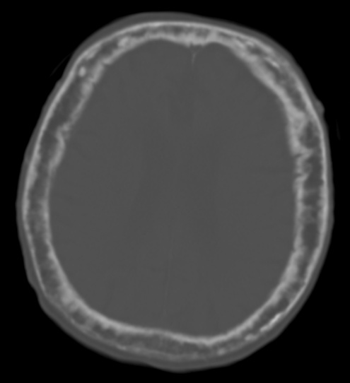


Paget disease of bone is a chronic focal disorder in which bone remodeling becomes excessive and disorganized. Osteoclasts resorb bone too rapidly, then osteoblasts replace it with structurally abnormal bone. The result is enlarged bone that may be mechanically weak or deformed despite increased bone mass. [1][2]
The disease is usually confined to one skeletal site in the monostotic form or affects multiple sites in the polyostotic form. Polyostotic lesions are typically asymmetrical rather than evenly distributed throughout the skeleton. The pelvis, femur, lumbar spine, skull, and tibia are the characteristic sites, so the clinical consequences depend strongly on which bone is involved. [1]
Pagetic remodeling evolves through three radiographic phases. The lytic phase reflects the early predominance of osteoclastic resorption and produces sharply defined osteolysis. The mixed phase combines resorption with abnormal new bone formation and is the most commonly observed phase. The sclerotic or blastic phase reflects later disorganized bone formation and produces cortical thickening, coarse trabeculae, enlargement, and deformity. These phases are not separate diseases or fixed time points because they form a continuous spectrum and may coexist within one bone. [1][3]
Radiographic phase and metabolic activity are related but not identical. An active pagetic lesion is undergoing increased remodeling and may cause pain or progressive enlargement, whereas an inactive or quiescent lesion represents residual abnormal bone after remodeling has substantially subsided. Such residual bone can remain enlarged and deformed even when current metabolic activity is low. Sclerotic or burnt-out lesions may show little uptake on metabolic imaging, so a striking deformity does not by itself prove that the lesion remains active. [1]
Many patients have no symptoms and are identified incidentally. When symptoms occur, bone pain is common, but pain may also arise from deformity or osteoarthritis near or distant from the pagetic bone. Disease may cause bowing or enlargement of bone, pathological fracture, nerve compression, hearing loss with skull involvement, and secondary osteoarthritis. [1][2][8]
| Phenotype | Typical skeletal sites | Radiographic appearance | Biochemical activity | Principal clinical implications |
|---|---|---|---|---|
| Monostotic disease | One site, often the pelvis, femur, spine, skull, or tibia | Changes are confined to one bone and may show any phase of Paget disease | Activity ranges from high during active remodeling to low after quiescence | Symptoms and complications are determined by the single involved bone; many cases are incidental or minimally symptomatic. [1] |
| Polyostotic disease | Multiple sites, commonly involving an asymmetrical combination of the pelvis, femur, spine, skull, and tibia | Multiple pagetic bones show lytic, mixed, or sclerotic change; different phases may coexist | Activity may differ between affected bones because lesions are not necessarily synchronized | The larger skeletal burden increases the opportunity for deformity, fracture, neurologic compression, hearing loss, and adjacent-joint osteoarthritis. [1][2] |
| Lytic phase | Early involvement of a long bone or the skull | Large, well-defined areas of osteolysis; a progressing long-bone lesion may form a flame-shaped or inverted-V advancing edge | Usually metabolically active because resorption predominates | Bone is being rapidly removed, so structural weakness and later deformity can develop. [1] |
| Mixed phase | Any pagetic site, especially the pelvis, long bones, spine, or skull | Osteolysis occurs with cortical and trabecular thickening; the pattern combines the cardinal features of Paget disease | Typically active because resorption and abnormal formation are both increased | This is the commonest radiographic presentation and may produce pain, enlargement, deformity, or mechanical complications. [1] |
| Sclerotic or blastic phase, including an inactive or quiescent residual lesion | Long bones, vertebrae, pelvis, or skull | Dense coarse bone with cortical thickening, loss of the corticomedullary distinction, enlargement, and deformity | Activity may remain high or may fall markedly when the lesion becomes burnt out; the residual deformity can persist after activity declines | Deformity and mechanical consequences may remain even when current remodeling is limited; fracture, nerve compression, hearing loss, or secondary osteoarthritis may reflect the old structural damage. [1][2] |
Pearl: Paget disease is best understood as a focal remodeling disorder whose current activity and residual skeletal damage must be considered separately. [1]
Who Develops Paget Disease: Age, Geography, Family History, and Genetic Susceptibility
- ▸Paget disease is uncommon before age 40 and usually diagnosed after age 50; a normal evaluation in midlife does not exclude later disease, especially when a first-degree relative is affected.
- ▸A pathogenic SQSTM1 variant occurs in up to 50% of familial cases, but a negative test does not remove the diagnosis or the possibility of inherited susceptibility, and carriers may remain disease-free for years.


is predominantly a disease of older adults. It is uncommon before age 40 and is usually diagnosed after age 50, although inherited disease can emerge earlier or later within the same family.[3] Men are affected slightly more often than women in epidemiologic studies.[3] This age pattern matters because a normal evaluation in midlife does not exclude later disease, especially when a first-degree relative is affected.
Occurrence varies sharply by geography and ancestry. Historically high-prevalence populations have clustered in the United Kingdom and Ireland, Australia, New Zealand, North America, and parts of Western Europe.[3][20] Prevalence has generally been lower in many Asian, African, and Scandinavian populations, although contemporary studies show that local population data can differ from broad historical patterns.[3][12][19] Therefore, ancestry and residence should modify clinical suspicion rather than determine it.
The incidence of has declined in several regions where the disease was previously common. Quebec showed a fall in standardized incidence from 0.77 per 1000 people in 2000–2001 to 0.28 per 1000 in 2019–2020, while studies from Britain and Spain also describe a continuing decline.[14][20][21] The decline cannot be explained by inherited susceptibility alone because genetic backgrounds change more slowly than disease incidence. Environmental exposure is therefore a plausible contributor, although the responsible exposure remains uncertain.[10][20][21]
Family history identifies a substantial but incomplete inherited component. A positive family history has been reported in approximately 15%–40% of patients, and first-degree relatives have a higher risk than people without affected relatives.[3][15] Familial disease often follows an autosomal-dominant pattern, meaning that one altered gene copy can confer susceptibility, but age-related penetrance is incomplete.[3][15] Thus, inheriting susceptibility does not guarantee disease, and the absence of a known family history does not exclude sporadic disease.
is the major known genetic association. This gene encodes p62, a cellular signaling protein, and pathogenic variants occur in up to 50% of familial cases while also occurring in a smaller proportion of apparently sporadic cases.[3][10] Most patients with therefore do not have an identifiable SQSTM1 variant, and a negative test does not remove the diagnosis or the possibility of inherited susceptibility.[3][15] Other rare genes and susceptibility loci account for additional familial or severe early-onset phenotypes, but the available evidence does not support describing every case as genetically explained.[3][10]
The clinical meaning of a pathogenic SQSTM1 variant is probabilistic rather than deterministic. In a long-term study of adult carriers who initially had negative bone scans, only a small proportion developed detectable disease during follow-up, and affected carriers generally developed disease later with less extensive involvement than their clinically affected parents.[10] This observation supports interaction between inherited susceptibility and changing environmental factors. Genetic testing is therefore most informative when a patient has a strong family history or when testing can guide surveillance of relatives, rather than as a universal test for every patient.[2][10]
| Epidemiologic feature | Observed pattern | Clinical relevance | Strength or limitation of evidence |
|---|---|---|---|
| Age | Predominantly older-adult disease; uncommon before 40 and rarely diagnosed before 50.[3] | Investigate unexplained disease in an older adult more readily than in a young adult. Consider an inherited or syndromic disorder when onset is unusually early.[3][25] | Consistent epidemiologic pattern, but age at diagnosis is influenced by incidental detection and access to imaging.[3][10] |
| Sex | Slight male predominance in classical epidemiologic studies.[3] | A woman can still have ; sex should not be used to rule it out.[3][19] | Reported across several cohorts, but the magnitude varies by population and age structure.[3][19] |
| Geography and ancestry | Highest historical prevalence has been reported in the United Kingdom and related settler populations in Australia, New Zealand, and North America, with additional clustering in Western Europe.[3][20] Many Asian, African, and Scandinavian populations have had low reported prevalence.[3] | Geographic background changes the prior probability of disease and should influence how actively incidental biochemical or radiographic abnormalities are investigated. | Strong geographic signal, but estimates vary with case ascertainment, diagnostic practice, migration, and underdiagnosis.[3][12][19][24] |
| Secular trend | Incidence has declined in Quebec and Spain, while Britain has also experienced reduced prevalence and less severe disease than historically.[14][20][21] | A patient from a historically high-prevalence region may have a lower current risk than older epidemiologic figures suggest. | Repeated regional observations support a real trend, but the causal environmental factor remains unproven.[10][14][20][21] |
| Family history | Approximately 15%–40% of patients report familial disease.[3][15] First-degree relatives have higher risk than people without affected relatives.[3] | Ask specifically about in parents, siblings, and children. A positive history supports targeted assessment of relatives.[2][3] | Family history underestimates familial disease because asymptomatic relatives may never have been diagnosed.[3] |
| SQSTM1 susceptibility | Pathogenic SQSTM1 variants are found in up to 50% of familial cases and in a smaller proportion of sporadic cases.[3][10] | A pathogenic result supports inherited susceptibility and may justify evaluation of at-risk relatives, but it does not prove that disease will develop.[2][10] | SQSTM1 is the major known association, yet many familial and sporadic cases remain unexplained.[3][15] |
| Penetrance of inherited susceptibility | Adult SQSTM1 carriers can remain scan-negative for years, and carriers who develop disease may do so later than their affected parents.[10] | Do not equate a pathogenic variant with inevitable disease or assume that a negative earlier scan permanently excludes future disease.[10] | Longitudinal evidence supports incomplete and age-dependent penetrance, but cohorts remain relatively small.[10] |
Why Pagetic Bone Remodels Abnormally: Osteoclasts, Osteoblasts, and SQSTM1 Signaling
- ▸Pagetic osteoclasts respond to lower concentrations of osteoclastogenic signals because mutant p62 reduces CYLD-mediated attenuation of TRAF6 signaling, leading to sustained NF-κB activation.
- ▸The resulting bone contains woven bone and mosaic lamellar architecture, so it appears densely mineralized on imaging but is mechanically weak, accounting for deformity and fracture risk.
- ▸Osteoclast hyperactivity is coupled to excessive osteoblast activity via coupling signals such as IGF1 and ephrinB2, so effective suppression of osteoclast activity also reduces abnormal bone formation.


The primary cellular abnormality in is the osteoclast. Pagetic osteoclasts are unusually numerous and large, and they contain excessive numbers of nuclei because their precursors fuse readily and respond to lower concentrations of osteoclastogenic signals.[26] They therefore resorb bone intensely before osteoblasts can replace it. This explains why the disease begins as a focal remodeling disturbance rather than as a generalized failure of bone formation.[13]
The central recruitment system is the – – axis. RANKL is a membrane-bound or soluble ligand produced mainly by osteoblast-lineage cells and osteocytes. It binds RANK on osteoclast precursors and recruits TRAF6, an adaptor protein that activates IKK and downstream . Osteoprotegerin, or OPG, is a soluble decoy receptor that binds RANKL before RANK can be engaged. Excessive RANKL signaling or inadequate OPG restraint therefore increases osteoclast differentiation and survival.[38]
RANK signaling converges on and . NF-kappa B is a transcription factor that enters the nucleus after IKK activation and permits expression of osteoclastogenic genes. NFATc1 is the lineage-defining transcription factor that consolidates this program by inducing genes required for osteoclast fusion and resorption. In Pagetic cells, the NFAM1 pathway can amplify calcium oscillations and activate PLCγ, calcineurin, and NFATc1, which helps explain the formation of hypermultinucleated osteoclasts and their increased resorptive activity.[36]
encodes p62, a scaffold protein that assembles signaling complexes downstream of cytokine receptors. Many Paget-associated p62 mutations disrupt its C-terminal ubiquitin-associated domain. This defect reduces recruitment of CYLD, a deubiquitinating enzyme that normally dampens TRAF6, RIP1, and NEMO signaling. NF-kappa B signaling therefore remains active after weaker receptor stimulation, so osteoclast precursors cross the differentiation threshold too easily.[26] The mutation does not by itself explain every lesion because experimental p62 models can increase osteoclast numbers without reproducing the complete Pagetic phenotype.[35]
Osteoclast hyperactivity is coupled to excessive osteoblast activity rather than followed by orderly repair. Pagetic osteoclasts release coupling signals such as and ephrinB2, which stimulate osteoblast differentiation through the EphB4 receptor and related pathways.[30] This is why effective suppression of osteoclast activity also reduces the abnormal rate of bone formation. The osteoblasts consequently lay down abundant bone rapidly, but the collagen is arranged irregularly instead of in normal lamellae.[13]
The resulting tissue contains woven bone and irregularly interleaved lamellar bone in a mosaic pattern. It expands the involved bone and can thicken the cortex, yet its disordered architecture transmits mechanical loads poorly. The same lesion can therefore appear densely mineralized while remaining structurally weak, which accounts for deformity and fracture risk.[26]
| Cellular event | Molecular mediator or pathway | Structural consequence | Clinical or imaging correlate |
|---|---|---|---|
| Osteoclast precursors are recruited too readily and form large multinucleated cells | RANKL activates RANK, TRAF6, IKK, NF-kappa B, and NFATc1; OPG normally restrains this signal.[36][38] | Excessive focal resorption creates irregular resorption surfaces | Osteolytic or mixed remodeling areas and increased local turnover.[28] |
| Pagetic osteoclasts respond to unusually low osteoclastogenic stimulus | Mutant p62 lowers CYLD-mediated attenuation of TRAF6 and related signaling.[26] | Resorption becomes prolonged and disproportionate to normal repair | Lesion activity can remain concentrated within a restricted skeletal site.[13] |
| Osteoclasts signal strongly to osteoblast-lineage cells | IL-6 increases osteoclast-derived IGF1, while IGF1 enhances ephrinB2–EphB4 coupling.[30] | Bone formation becomes excessive but poorly coordinated | Rapid sclerosis and bony expansion follow the earlier resorptive phase.[26] |
| Osteoblasts repair the resorbed surface at high speed | Coupling signals increase osteoblast differentiation and matrix production.[31] | Woven bone and mosaic lamellar bone replace orderly lamellar architecture | Enlarged bone may be dense on radiographs yet mechanically weak.[26][28] |
A paramyxovirus-related trigger remains a proposed cofactor rather than an established cause. Measles virus, respiratory syncytial virus, and canine distemper virus have each been reported in some studies, but other investigations failed to detect these viruses in Pagetic cells. A large antibody study also found no evidence that Paget disease is associated with persistent measles or other paramyxovirus infection.[42] Experimental measles nucleocapsid protein models can reproduce Pagetic-like osteoclasts and lesions, especially when combined with mutant p62, so viral signaling may help explain disease expression in a genetically susceptible bone without proving that infection initiates human disease.[35]
How Paget Disease Presents: Bone Pain, Deformity, Joint Disease, and Neurologic Features
- ▸Active Paget disease accounts for only 14.7% of painful presentations, while osteoarthritis at a distant site accounts for 44.1%.
- ▸A new inability to walk or a sudden increase in pain warrants assessment for fracture, nerve compression, or another superimposed disorder rather than assuming uncomplicated Paget disease.
- ▸Hearing loss from skull involvement can be sensorineural, conductive, or mixed and should not be dismissed as ordinary age-related hearing loss.


has a broad clinical range, from an incidental finding to disabling mechanical or neurologic disease. Pain is the commonest symptom, yet many patients have no skeletal symptoms when the diagnosis is made. [44] Pain therefore requires anatomical localisation rather than automatic attribution to active pagetic remodeling. [8]
Pagetic bone pain may come from active remodeling because rapid turnover can stimulate local nociceptive pathways. It may also arise from a pathological fracture, deformity, adjacent-joint , or nerve compression. [8] In a contemporary clinical study, active Paget disease accounted for only 14.7% of painful presentations, whereas osteoarthritis at a distant site accounted for 44.1%. [8] A painful pagetic site can therefore be metabolically quiet, while active disease can be painless. [8]
Local warmth occurs because blood flow increases through affected pagetic bone. [1] The same abnormal remodeling can enlarge and weaken bone, producing thickened contours, bowing of a long bone, or limb shortening. [1][8] Bowing changes the mechanical axis and may produce an antalgic or broad-based gait because loading becomes painful or inefficient. [44] A new inability to walk or a sudden increase in pain warrants assessment for fracture, nerve compression, or another superimposed disorder rather than assuming uncomplicated Paget disease. [53]
Skull involvement may enlarge the calvarium and produce frontal bossing because the outer skull contours expand. Patients may report a larger hat size, scalp discomfort, headache, or facial asymmetry. [56] Skull disease can also affect the temporal bone and cranial nerves, causing hearing loss, tinnitus, vertigo, or less commonly visual symptoms from optic-nerve compression. [9][5] Hearing impairment may be sensorineural, conductive, or mixed, so symptoms should not be dismissed as ordinary age-related hearing loss. [9]
Spinal involvement causes axial pain through abnormal vertebral structure, deformity, fracture, or narrowing around neural elements. Nerve-root compression may cause radicular pain, paraesthesia, weakness, or reflex change, while spinal stenosis can produce claudication and progressive walking difficulty. [44][53] Cranial neuropathies likewise reflect compression or distortion around foramina and the skull base. [1] Neurologic symptoms that are focal, progressive, or acute require evaluation for a structural complication or an alternative lesion. [53]
Pagetic deformity increases abnormal mechanical loading across nearby joints, which can lead to secondary of the hip or knee. [1] Hip disease typically causes groin or buttock pain with reduced internal rotation, whereas knee disease causes joint-line pain and pain during weight-bearing. These patterns support joint disease when symptoms are movement-related and imaging confirms osteoarthritis at the painful joint. [8] The painful joint may be adjacent to pagetic bone or distant from it, so the distribution of osteoarthritis does not by itself prove that Paget disease is the pain source. [8]
Jaw or maxillofacial involvement can produce facial asymmetry, enlargement of the maxilla or mandible, altered occlusion, and dental difficulty. [1][25] Early tooth-root resorption and hearing loss are especially characteristic of rare inherited expansile osteolytic disorders that can resemble or overlap with Paget-spectrum disease, particularly when symptoms begin unusually early. [25] In an older adult with skull or jaw disease, ask about chewing difficulty, loose or displaced teeth, facial change, hearing symptoms, and visual symptoms because these findings localise clinically important craniofacial involvement. [1][25]
| Presentation | Likely anatomic site | Possible mechanism | Findings that support active Paget disease | Important alternative diagnosis |
|---|---|---|---|---|
| Deep focal bone pain | Pelvis, femur, tibia, spine, or another pagetic bone | Active remodeling, deformity, fracture, or local structural stress | Pain co-localises with a known pagetic site and accompanies new local symptoms or warmth | Osteoarthritis, metastatic bone disease, infection, stress fracture, or unrelated regional pain [8][53] |
| Local warmth | Affected long bone, pelvis, or skull | Increased blood flow through metabolically active pagetic bone | Warmth is directly over an enlarging or painful pagetic site | Cellulitis, inflammatory arthritis, trauma, or another hypervascular bone lesion [1] |
| Enlargement or thickening of bone | Skull, pelvis, femur, tibia, or other involved bone | Excessive disorganised formation expands the affected bone | Progressive local enlargement corresponds to a pagetic lesion on skeletal imaging | Fibrous dysplasia, chronic infection, metastatic disease, or an expansile tumour [1][45] |
| Bowing, limb shortening, or gait disturbance | Femur and tibia most often | Weak enlarged bone deforms under load and changes limb alignment | Visible deformity, altered limb axis, local pain, or worsening walking mechanics at the involved limb | Osteoarthritis, previous fracture, neuromuscular disease, or spinal stenosis [1][8][44] |
| Skull enlargement or frontal bossing | Calvarium, especially the frontal skull | Expansion and thickening of pagetic skull bone | Progressive head enlargement or frontal contour change with other skull symptoms | Acromegaly, fibrous dysplasia, osteoma, or another cranial bone lesion [1][56] |
| Hip pain or knee pain | Hip or knee adjacent to deformed pelvis, femur, or tibia | Secondary osteoarthritis from altered mechanical loading | Movement-related joint pain with radiographic osteoarthritis at the symptomatic joint | Primary osteoarthritis, inflammatory arthritis, meniscal disease, or referred spinal pain [1][8] |
| Spinal pain | Lumbar, thoracic, or cervical vertebrae | Vertebral deformity, fracture, abnormal loading, or neural compression | Focal spinal pain corresponds to pagetic vertebral involvement and may coexist with deformity or neurologic signs | Metastasis, infection, degenerative spondylosis, compression fracture, or myeloma [1][53] |
| Radiculopathy or spinal stenosis | Pagetic spine and adjacent neural foramina or canal | Nerve-root or canal compression from enlarged or deformed bone | Dermatomal pain, sensory change, weakness, claudication, or progressive gait limitation with spinal involvement | Disc disease, degenerative stenosis, epidural tumour, or cauda equina compression from another cause [44][53] |
| Hearing loss, tinnitus, or vestibular symptoms | Temporal bone, otic capsule, or skull base | Bony remodeling may affect the cochlea or auditory pathways and may disturb vestibular structures | Symptoms occur with skull involvement and may include sensorineural, conductive, or mixed hearing loss | Presbycusis, otosclerosis, Ménière disease, vestibular schwannoma, or ototoxicity [9][54] |
| Visual or other cranial neuropathy | Skull base and cranial-nerve foramina | Compression or distortion of cranial nerves by enlarged pagetic bone | Focal cranial-nerve deficit with skull-base involvement | Intracranial tumour, vascular compression, inflammatory neuropathy, or metastatic disease [1][5] |
| Dental or maxillofacial change | Maxilla, mandible, or adjacent facial bones | Jaw expansion can alter facial contour, occlusion, and tooth position | Facial enlargement or dental change accompanies pagetic craniofacial involvement | Fibrous dysplasia, giant-cell lesion, odontogenic tumour, or inherited expansile osteolysis [1][25] |
Confirming Paget Disease: Alkaline Phosphatase, Bone-Turnover Markers, and Differential Diagnosis
- ▸A normal total alkaline phosphatase does not exclude Paget disease; 42% of cases have normal t-ALP, particularly in monostotic or inactive disease.
- ▸When t-ALP is normal or confounded by liver disease, use bone-specific alkaline phosphatase (B-ALP) or procollagen type 1 intact N-terminal propeptide (PINP) as more specific markers of bone turnover.
- ▸Before initiating potent antiresorptive therapy, obtain serum calcium, phosphate, creatinine, 25-hydroxyvitamin D, and liver tests; measure PTH if calcium is abnormal or if vitamin D deficiency or renal disease is present.


A diagnosis of requires concordance between characteristic skeletal imaging and evidence of increased bone remodeling. Total alkaline phosphatase (t-ALP) is the usual first biochemical test because it is widely available and reflects osteoblast activity. Interpret it with liver tests because t-ALP also rises in hepatobiliary disease; an isolated elevation therefore supports a bone source but does not by itself establish Paget disease. [1][2]
A normal t-ALP does not exclude limited or inactive disease. In one population-based study, 42% of Paget cases had normal t-ALP. Monostotic disease may produce a normal or only modest elevation because the affected bone contributes little to total skeletal enzyme release. Burnt-out sclerotic lesions may also show little current remodeling, so the radiographic abnormality persists after biochemical activity has fallen. [1][2]
Use a bone-specific marker when t-ALP is normal despite convincing imaging or when liver disease makes total enzyme interpretation unreliable. Bone-specific alkaline phosphatase (B-ALP) measures osteoblast-derived alkaline phosphatase more directly. Procollagen type 1 intact N-terminal propeptide (PINP) reflects newly synthesized type I collagen and is often particularly useful in limited disease or coexisting liver disease. Neither marker is disease-specific, so an abnormal result still requires compatible skeletal imaging. [1][2]
Serum C-terminal telopeptide (CTX) and urinary CTX reflect type I collagen breakdown and therefore osteoclastic resorption. These markers can support active disease when formation markers are equivocal, and all major turnover markers correlate with scintigraphic activity. CTX is affected by sampling conditions and renal clearance, while urinary markers have greater biological and preanalytical variability; use a consistent fasting morning sample when CTX is needed and interpret the result with renal function. [1][2][57]
Before potent , obtain serum calcium, phosphate, creatinine, 25-hydroxyvitamin D, and liver tests. Measure when calcium is abnormal or when vitamin D deficiency or renal disease could be causing secondary hyperparathyroidism. This baseline matters because severe active disease can increase skeletal calcium uptake, while antiresorptive therapy can precipitate hypocalcemia when vitamin D or calcium supply is inadequate. [1][2][56]
Use imaging to resolve biochemical uncertainty rather than escalating markers indefinitely. Plain radiographs of the suspected site are usually diagnostic when they show the characteristic combination of bone enlargement, cortical thickening, coarse trabeculae, sclerosis, or deformity. No single radiographic feature is pathognomonic, so an isolated lytic or sclerotic focus requires a broader differential. A radionuclide bone scan maps metabolically active skeletal involvement, but a sclerotic burnt-out lesion may show little or no uptake. [1][2]
| Test or mimic | Expected result or imaging clue | Usefulness | Major limitation | Next step |
|---|---|---|---|---|
| Total alkaline phosphatase with liver tests | Isolated t-ALP elevation supports increased bone turnover. Normal t-ALP can occur in limited monostotic disease or inactive disease. [1][2] | First-line biochemical assessment of activity and a practical baseline for follow-up. | Liver disease can elevate t-ALP and a normal value does not exclude Paget disease. [1][2] | Confirm the skeletal source with B-ALP or PINP and correlate with targeted radiographs. |
| B-ALP | Increased when osteoblast activity is increased, including in Paget disease. [1][2] | Useful when t-ALP is confounded by liver disease or is unexpectedly normal. | It still reflects bone formation rather than a Paget-specific process. [1][2] | Obtain compatible radiographs and consider bone scanning to define active sites. |
| PINP | Increased bone formation marker that may detect activity in limited disease. [2] | Helpful when liver disease limits t-ALP interpretation and for detecting biochemical recurrence. [2] | Availability and cost are less favorable than t-ALP. [1] | Use as a corroborating marker rather than as a stand-alone diagnosis. |
| Serum or urinary CTX | Increased resorption marker may accompany active Paget disease. [1][2] | Adds information when formation and resorption may be discordant. | CTX varies with fasting status and renal function. Urinary assays have greater biological variation. [57] | Standardize collection and interpret alongside formation markers and imaging. |
| Fibrous dysplasia | A lytic lesion may resemble early Paget disease but usually lacks the progressive pagetic combination of cortical thickening and whole-bone expansion. [1] | Important alternative for an isolated lucent or expansile lesion. | Imaging overlap can be substantial in a monostotic lesion. [1] | Use CT for matrix and cortical characterization and biopsy if imaging remains atypical. [1] |
| Osteomalacia | Calcium or phosphate disturbance and secondary hyperparathyroidism may point toward defective mineralization rather than focal Paget remodeling. [1][56] | Prevents misattributing diffuse high turnover to Paget disease. | Biochemical values can be normal early or after partial correction. | Check 25-hydroxyvitamin D, phosphate, calcium, creatinine, and PTH, then reassess the skeletal findings. |
| Hyperparathyroidism | Hypercalcemia or an inappropriately high PTH suggests a systemic high-turnover disorder. [1][56] | Identifies a potentially reversible cause of raised ALP and bone turnover. | Mild primary disease can have normal calcium, and secondary disease may coexist with Paget disease. [56] | Repeat calcium with PTH and evaluate vitamin D and renal function before assigning the abnormality to Paget disease. |
| Metastatic bone disease | Multifocal lesions or focal lysis may mimic Paget disease, while bone scan uptake is not specific because metastases can also accumulate tracer. [1] | Essential when lesions are atypical, painful, rapidly progressive, or discordant with the usual pagetic distribution. | A bone scan alone cannot reliably distinguish metastases from Paget disease. [1] | Review prior imaging and cancer history, use cross-sectional imaging, and biopsy a suspicious lesion. [53][61] |
| Chronic recurrent multifocal osteomyelitis | Multifocal inflammatory bone lesions may cause pain and increased turnover but usually have an inflammatory or recurrent clinical pattern rather than classic pagetic expansion. | Keeps infection-related and sterile inflammatory bone disease in the differential. | Clinical and imaging overlap may be considerable. | Obtain inflammatory markers and targeted MRI; pursue microbiologic or histologic evaluation when infection or malignancy remains plausible. |
| Osteosarcoma | A new aggressive lytic or mixed lesion with cortical destruction, soft-tissue mass, or rapidly worsening pain is atypical for uncomplicated Paget disease. [1][61] | Detects rare malignant transformation or an unrelated primary bone tumor. | A pre-existing pagetic bone can obscure the tumor's margins. | Arrange urgent oncologic imaging and biopsy before definitive treatment. [1][61] |
| Giant-cell tumor | An osteoclast-rich expansile lesion can arise in pagetic bone and may resemble a conventional giant-cell tumor. [1][63] | Explains new focal deterioration within apparently established Paget disease. | Imaging and histology may require molecular and clinicopathologic correlation. [63] | Refer for specialist bone-tumor review and obtain tissue diagnosis. |
| Fracture healing | A healing fracture can produce focal sclerosis and increased turnover or scan uptake. [1][60] | Prevents mislabeling a stress or insufficiency fracture as active Paget disease. | Deformed pagetic bone is itself prone to stress fracture, so both conditions may coexist. [1] | Obtain dedicated radiographs and CT or MRI when pain is acute or radiographs are equivocal. |
| Degenerative sclerosis | Osteoarthritis or other degenerative change can cause focal sclerosis and increased scintigraphic uptake near a joint. [1] | Helps separate adjacent-joint disease from an underlying pagetic bone lesion. | Degenerative disease may coexist with Paget disease and may explain pain without explaining bone enlargement. [1][2] | Localize symptoms and compare the entire involved bone on radiographs rather than interpreting a single sclerotic focus. |
When biochemical results and imaging disagree, do not label the patient solely from an elevated marker or a positive scan. Review the radiographs for whole-bone expansion and the characteristic mixed pattern, then use CT or MRI for an aggressive, structurally complex, or fracture-related lesion. Perform a biopsy when imaging remains atypical or when osteosarcoma, giant-cell tumor, metastatic disease, or another neoplasm cannot be excluded. [1][60][61]
Pearl: A normal t-ALP lowers the probability of extensive active Paget disease but does not rule out a small monostotic lesion or a metabolically inactive sclerotic lesion; in that situation, the radiographic pattern and targeted imaging carry greater diagnostic weight. [1][2]
Mapping Pagetic Lesions: Radiographs, Bone Scintigraphy, CT, MRI, and Biopsy
- ▸Plain radiography is the primary diagnostic test and should include the entire suspected bone rather than only the painful segment to distinguish focal Paget disease from an aggressive lesion.
- ▸Bone biopsy is not routine for a typical radiographic pattern; perform it when imaging is atypical, progression is unexplained or rapid, or sarcoma cannot be excluded.
- ▸Use MRI when neural compression is suspected or when evaluating for sarcomatous transformation, as diffuse marrow replacement, cortical erosion, and soft-tissue extension are warning signs.
is the primary diagnostic test because Pagetic bone usually has a recognizable whole-bone pattern. [45] The appearance changes with remodeling phase and may vary along the same bone.
- In the early osteolytic phase, resorption produces a sharply marginated radiolucency. In long bones this may advance as a tapering blade-of-grass or flame-shaped lucency, especially in the femur and tibia. [45][61]
- In the mixed phase, new bone formation thickens the cortex and makes the trabeculae coarse. The involved bone expands and may bow because abnormal remodeling enlarges the bone without restoring normal mechanical structure. [45][61]
- In the late sclerotic phase, the bone becomes densely mineralized and remains enlarged or deformed. In the skull, patchy thickening creates the classic cotton-wool appearance, while long bones may show marked cortical thickening and bowing. [45]
Radiographs should include the entire suspected bone rather than only the painful segment. This approach shows whether a lucency continues into a typical Pagetic transition and distinguishes focal Paget disease from an aggressive lesion with cortical destruction or a soft-tissue mass. [61]
maps metabolically active Pagetic bone across the skeleton. Increased tracer uptake reflects increased local osteoblastic activity, so scintigraphy is useful for detecting clinically silent sites and for estimating disease distribution. [44][67] It is not specific for Paget disease and may show little uptake in an inactive or burnt-out lesion. Therefore, obtain targeted radiographs of every scintigraphically abnormal site because the scan answers where activity is present, whereas radiographs establish whether the bone has the characteristic Pagetic structure. [44][45]
is not required to confirm a typical lesion, but it clarifies anatomy when plain films are difficult to interpret. Use it for complex skull-base or spinal anatomy, precise cortical assessment, suspected fracture, and operative planning. [45] CT is particularly useful when a radiograph suggests Paget disease but the cortex appears permeated or destructed, because those findings may indicate another process. [61]
answers a different question from CT. Use it when neural compression is suspected or when symptoms suggest spinal or skull-base complications. MRI also evaluates marrow replacement and soft-tissue extension, which are warning signs for sarcomatous transformation or another tumor rather than uncomplicated Paget disease. [61] New focal pain, rapidly progressive symptoms, or neurologic deterioration should lower the threshold for MRI because a superimposed fracture or malignancy can occur within apparently pagetic bone. [53][68]
is not routine when the radiographic pattern is typical and the clinical course is concordant. [61] Perform an image-guided or surgical biopsy when imaging is atypical, progression is unexplained or unusually rapid, or sarcoma and another tumor cannot be excluded. [61] A soft-tissue mass, permeative cortical destruction, or extensive marrow replacement is especially concerning because these findings fall outside the usual imaging spectrum of Paget disease. [61] Biopsy then changes management by providing histologic confirmation and directing oncologic or other disease-specific treatment rather than empiric Paget care.
| Modality | Best indication | Characteristic Paget findings | Limitations | Effect on management |
|---|---|---|---|---|
| Plain radiography | Initial diagnosis and characterization of the entire involved bone | Early well-defined osteolysis; blade-of-grass or flame-shaped lucency; later cortical thickening, coarse trabeculation, expansion, sclerosis, cotton-wool skull, and bowing [45][61] | May underestimate total skeletal distribution and cannot reliably distinguish current activity from residual deformity [44][45] | Usually establishes the diagnosis and identifies sites needing further characterization [44][45] |
| Technetium-labeled diphosphonate bone scintigraphy | Whole-skeleton mapping of metabolically active disease | Increased uptake in active Pagetic sites [67] | Uptake is not disease-specific and may be low in burnt-out lesions [44][45] | Determines where to obtain targeted radiographs and reveals clinically silent skeletal involvement [44][67] |
| CT | Complex anatomy; cortical detail; suspected fracture; surgical planning | Cortical thickening, trabecular coarsening, expansion, and detailed lytic or sclerotic architecture [45][61] | Involves ionizing radiation and provides less sensitive assessment of marrow and neural soft tissues than MRI | Defines structural risk and operative anatomy; atypical cortical destruction prompts MRI or biopsy [45][61] |
| MRI | Neural compression; marrow abnormality; suspected sarcomatous transformation or another tumor | Abnormal marrow signal may occur in Paget disease; diffuse marrow replacement, cortical erosion, and soft-tissue extension are atypical warning findings [61] | Pagetic marrow signal can overlap with tumor or other marrow disorders, so MRI may not provide a definitive diagnosis [61] | Detects complications and determines whether tissue diagnosis or urgent decompression is needed [45][61] |
| Bone biopsy | Atypical imaging or unresolved concern for sarcoma or another tumor | Histologic confirmation when imaging cannot establish typical Paget disease [61] | Invasive and unnecessary for a characteristic uncomplicated lesion [61] | Separates Paget disease from malignancy or another tumor and directs definitive treatment [61] |
Assessing Activity, Extent, Severity, and Treatment Indications in Paget Disease
- ▸Disease-modifying treatment is clearly indicated for pagetic bone pain and for active disease at a site that threatens neurologic function or major mechanical integrity.
- ▸For asymptomatic patients, treat only when the lesion is active and high risk, or when selected orthopedic surgery is planned through pagetic bone; otherwise, document the structural and metabolic phenotype and monitor.
- ▸Use total alkaline phosphatase when liver tests are normal; if normal despite convincing imaging, or if liver disease confounds interpretation, use bone-specific alkaline phosphatase or PINP.
Active means ongoing remodeling rather than merely abnormal bone shape. Assess activity by combining symptoms with a bone-turnover marker and active uptake on . A persistently enlarged or sclerotic bone may remain abnormal after remodeling has quieted, so radiographic severity alone does not prove current activity. [1][76]
Localize pain before attributing it to Paget disease. Pagetic bone pain supports treatment when it arises from an active lesion and has no better explanation, whereas pain from adjacent , fracture, deformity, or nerve compression requires separate evaluation. This distinction matters because metabolic activity and pain correlate imperfectly. [76][1]
Use total when liver tests are normal and interpret it as a systemic estimate of skeletal turnover. If total alkaline phosphatase is normal despite convincing imaging, or if liver disease confounds interpretation, use bone-specific alkaline phosphatase or PINP, which is the procollagen type 1 N-terminal propeptide. A normal marker does not exclude limited monostotic disease or an inactive sclerotic lesion. [1][77]
Obtain a whole-skeleton radionuclide scan after radiographic confirmation to map the disease. Intense uptake indicates sites with increased remodeling, including clinically silent lesions, while absent uptake may indicate a burnt-out lesion rather than absence of structural Paget disease. Because uptake is not specific, obtain targeted radiographs for abnormal scan sites and resolve discordance with focused imaging. [1][76]
Extent and location determine risk more reliably than a single biochemical value. Skull lesions can threaten hearing or other cranial-nerve function, spinal lesions can narrow the canal or compress neural structures, and lesions in weight-bearing long bones can deform or fracture under load. Periarticular lesions increase abnormal joint loading and therefore the risk of secondary osteoarthritis. [76][44][1]
There is no TNM-style stage and no universally accepted activity or severity score for Paget disease. Describe burden instead by the number of involved bones, the proportion of each bone affected, the presence of deformity or fracture, biochemical activity, scintigraphic activity, symptoms, and the anatomic consequences of the lesion. This multidimensional description prevents a normal alkaline phosphatase from falsely reassuring clinicians when a high-risk site remains structurally involved. [76][75]
| Assessment domain | Measure | Interpretation | High-risk finding | Treatment consequence |
|---|---|---|---|---|
| Symptoms | Localized pagetic bone pain | Pain supports clinically significant disease when the painful site matches an active lesion | Persistent pain attributable to Paget disease | Offer disease-modifying antiresorptive therapy to relieve pagetic pain [77][1] |
| Neurologic function | Cranial-nerve and neurologic examination with symptom-directed CT or MRI | Symptoms indicate an anatomic consequence rather than biochemical activity alone | Skull-base hearing or visual symptoms; spinal stenosis or progressive weakness | Treat active disease promptly and coordinate structural evaluation because neurologic function is threatened [76][77] |
| Bone turnover | Total alkaline phosphatase with liver tests; use bone-specific alkaline phosphatase or PINP when needed | Elevation supports active remodeling but is not disease-specific | Markedly increased or rising turnover with a lesion at a mechanically or neurologically important site | Supports treatment and provides a baseline for follow-up [1][77] |
| Scintigraphic activity | Whole-skeleton technetium-labeled diphosphonate scan | Uptake identifies metabolically active skeletal sites and maps silent disease | Intense uptake in the skull, spine, weight-bearing long bone, or periarticular bone | Supports treatment when symptoms or future complication risk justify it; absent uptake does not erase structural risk [1][76] |
| Structural burden | Whole-bone radiographs | Expansion, cortical thickening, coarse trabeculae, sclerosis, and deformity show accumulated skeletal damage | Bowed or fissured weight-bearing long bone; enlarged skull; deformed or compressed vertebra | Raises concern for fracture, neurologic compromise, or mechanical failure even when turnover is low [1][76] |
| Distribution | Number of bones and extent within each involved bone | Monostotic disease can still be high risk if it involves a critical site; polyostotic disease increases skeletal burden | Extensive polyostotic disease or a lesion spanning a major load-bearing segment | Weighs toward treatment when combined with activity, symptoms, or site-specific risk [1][77] |
| Periarticular involvement | Relationship of the pagetic lesion to the hip, knee, or other major joint | Abnormal bone geometry can increase joint loading | Active disease adjacent to a major weight-bearing joint with mechanical symptoms | Supports treatment when Paget disease contributes to pain or threatens mechanical function [1][76] |
| Operative planning | Site activity and vascularity before selected orthopedic surgery | Active pagetic bone can complicate reconstruction and increase operative bleeding | Planned surgery through active pagetic bone | The Endocrine Society advises bisphosphonate treatment before surgery on pagetic bone [77] |
Disease-modifying treatment is clearly indicated for pagetic bone pain and for active disease at a site that threatens neurologic function or major mechanical integrity. The Endocrine Society guideline from 2014 suggests a bisphosphonate for most patients with active disease who are at risk for future complications, with a conditional recommendation because evidence for preventing complications remains limited. [77]
Selected asymptomatic patients may also merit treatment when active disease affects the skull, spine, a weight-bearing long bone, or bone adjacent to a major joint. The decision should reflect the lesion’s activity, extent, deformity, and plausible future consequence rather than an isolated laboratory threshold. The 2019 guideline developed with the UK Paget’s Association, the European Calcified Tissue Society, and the International Osteoporosis Foundation and the Endocrine Society guideline do not establish a single universal treatment threshold; reviews explicitly describe disagreement about treatment indications. [76][75][78]
Do not automatically treat every asymptomatic patient with biochemical activity. The PRISM and PRISM-EZ studies found that intensive treatment aimed at normalizing alkaline phosphatase was no more effective than symptom-directed treatment for preventing complications, while evidence that early treatment prevents fracture, osteoarthritis, hearing loss, or deformity remains inadequate. [82][1] Treat an asymptomatic patient when the lesion is active and high risk, or when selected orthopedic surgery is planned through pagetic bone; otherwise, document the structural and metabolic phenotype and monitor it. [77][82]
Pearl: Active Paget disease is a metabolic diagnosis with an anatomic consequence. Treat the patient whose active lesion causes pain or threatens function, not the patient whose bone is merely permanently enlarged or sclerotic.
Disease-Modifying Therapy: Zoledronic Acid and Other Antiresorptive Options
- ▸Zoledronic acid 5 mg intravenously as a single infusion is the treatment of choice for active Paget disease when no contraindication exists.
- ▸Correct vitamin D deficiency and hypocalcemia before zoledronic acid infusion, and do not give the drug in severe renal impairment or untreated hypocalcemia.
- ▸Do not treat an isolated biochemical abnormality solely to normalize a laboratory value when the patient has no relevant clinical risk.
suppresses the excessive remodeling that sustains active . The Endocrine Society recommends a bisphosphonate for most patients with active disease who face future complications and suggests treatment before surgery on pagetic bone. [77] This strategy can normalize turnover and reduce disease-related pain, but it cannot reliably reverse established bowing, enlargement, fracture deformity, or adjacent-joint because those structural changes reflect bone that has already remodeled abnormally. [80]
The preferred regimen is 5 mg intravenously as one infusion. The Endocrine Society specifically suggests this single dose as the treatment of choice when no contraindication exists. [77] A single infusion produced biochemical response in 38 of 39 patients over a mean follow-up of 56.5 months, and another cohort found sustained response after treatment over long-term follow-up. [90] Reports describe normalization of alkaline phosphatase for up to 6.5 years, which explains why zoledronic acid is more durable than repeated oral courses. [66] Do not repeat the dose simply because the patient has residual deformity or osteoarthritis; reserve further treatment for demonstrably recurrent active disease or a new clinical indication.
Before infusion, measure serum creatinine and calculate renal function. Also measure calcium, phosphate, and 25-hydroxyvitamin D because potent osteoclast suppression can expose impaired mineral homeostasis and precipitate hypocalcemia. [76] Correct vitamin D deficiency and hypocalcemia before treatment, and do not give zoledronic acid in severe renal impairment or untreated hypocalcemia. Confirm adequate hydration and avoid concurrent nephrotoxins when possible. A common first-infusion acute-phase reaction causes fever, myalgia, or influenza-like symptoms; these reactions occur in up to 40%–70% of first intravenous aminobisphosphonate infusions. [92] Vitamin D repletion with cholecalciferol 50,000 IU orally once weekly for 8 weeks before infusion reduced this risk in an interventional study. [92]
Discuss uncertainty about rather than presenting it as an established contraindication. A prior atrial-fibrillation signal has not established a causal relationship specific to zoledronic acid, so assess cardiovascular risk and use an alternative when the anticipated benefit is marginal. Examine for active dental infection and complete invasive dental work when feasible before treatment because rare has been associated with potent antiresorptive exposure. Ask about thigh or groin pain during subsequent care because persistent prodromal pain may indicate an ; investigate promptly rather than automatically repeating bisphosphonate therapy.
Oral bisphosphonates remain effective when intravenous therapy is unsuitable, but they require substantially more patient participation. The patient must take the tablet fasting with plain water, remain upright for at least 30 minutes, and avoid food or other medicines during the absorption window. This is why dysphagia, severe reflux, esophagitis, inability to sit or stand upright, and poor adherence favor intravenous treatment. Alendronate and risedronate provide better biochemical suppression and longer remission than etidronate, although direct comparative evidence between alendronate and risedronate is limited. [88] In real-world treatment, 3–4 months of weekly osteoporosis-dose therapy produced a median remission of 8.8 months, which illustrates the generally lower durability of oral therapy than the usual single zoledronic acid infusion. [91]
is now mainly a specialist option for resistant disease or when other bisphosphonates cannot be used. High-dose courses can improve symptoms and suppress turnover, but complete biochemical remission is difficult to achieve in resistant cases. [93] is less potent and less durable than bisphosphonates, so use it only when bisphosphonates are unsuitable or cannot be tolerated. A practical regimen is salmon calcitonin 100 IU subcutaneously once daily for up to 6 months, with treatment stopped earlier if biochemical response is inadequate; nausea, flushing, and tachyphylaxis limit prolonged use. Calcitonin and bisphosphonates both have documented efficacy, but zoledronic acid is by far the most effective treatment. [66]
| Therapy | Regimen | Biochemical efficacy | Durability | Adverse effects | Contraindications | Preferred clinical use |
|---|---|---|---|---|---|---|
| Zoledronic acid | 5 mg IV once over at least 15 minutes; do not schedule routine repeat dosing | Normalizes alkaline phosphatase in about 97% in a prospective cohort [90] | Often several years; normalization has been reported for up to 6.5 years [66] | Acute-phase reaction; hypocalcemia; renal toxicity; rare osteonecrosis of the jaw and atypical femoral fracture | Severe renal impairment; hypocalcemia; uncorrected vitamin D deficiency; untreated dental infection requiring urgent invasive treatment | First choice for active disease when renal and mineral safety criteria are met; the Endocrine Society recommends a single 5-mg dose [77] |
| Alendronate | 40 mg orally once daily for 6 months; take fasting with water and remain upright for at least 30 minutes | Effective suppression of alkaline phosphatase; stronger and more durable than etidronate [88] | Usually months rather than years; weekly osteoporosis-dose courses had median remission of 8.8 months [91] | Esophageal irritation; dyspepsia; hypocalcemia; rare osteonecrosis of the jaw and atypical femoral fracture | Esophageal emptying disorders; inability to remain upright; hypocalcemia; severe renal impairment | Oral alternative when infusion is unsuitable and gastrointestinal tolerance and adherence are reliable |
| Risedronate | 30 mg orally once daily for 2 months; take fasting with water and remain upright for at least 30 minutes | 71.1% normalized alkaline phosphatase after the first treatment cycle in postmarketing surveillance [58] | Relapse reached 12.9% by 40 weeks after the first cycle [58] | Gastrointestinal symptoms; hypocalcemia; rare osteonecrosis of the jaw and atypical femoral fracture | Esophageal emptying disorders; inability to remain upright; hypocalcemia; severe renal impairment | Oral alternative when a shorter course is preferred and gastrointestinal tolerance is acceptable |
| Etidronate | 5–10 mg/kg orally once daily for 6 months; take separately from calcium and food | Less potent biochemical suppression than newer oral bisphosphonates [88] | Generally shorter remission than alendronate or risedronate [88] | Gastrointestinal symptoms; impaired mineralization at excessive or prolonged exposure; hypocalcemia | Hypocalcemia; osteomalacia; severe renal impairment | Rarely used because newer bisphosphonates provide greater suppression and better durability [88] |
| Pamidronate | 30 mg IV daily for 3 consecutive days; specialist protocols may use repeated courses | Can improve resistant disease, but high-dose treatment normalized alkaline phosphatase in only one of five resistant patients [93] | Variable and often incomplete in resistant disease [93] | Acute-phase reaction; hypocalcemia; renal toxicity; rare osteonecrosis of the jaw | Severe renal impairment; hypocalcemia; uncorrected vitamin D deficiency | Specialist rescue treatment for resistant disease when standard therapy is unsuitable |
| Salmon calcitonin | 100 IU SC once daily for up to 6 months; stop if response is inadequate | Suppresses turnover but is less potent than bisphosphonates [66] | Shorter and less reliable than bisphosphonate remission [66] | Nausea; flushing; injection reactions; tachyphylaxis | Hypocalcemia; hypersensitivity to calcitonin; prolonged use when no biochemical response occurs | Temporary alternative when bisphosphonates are contraindicated or not tolerated |
The Endocrine Society and the 2019 international clinical guideline agree that zoledronic acid is the standard biochemical treatment, but treatment thresholds differ. The Endocrine Society suggests treating most active patients at risk of complications, whereas the 2019 guideline emphasizes symptom-directed treatment because PRISM and PRISM-EZ found no preventive advantage from intensive alkaline-phosphatase normalization in otherwise uncomplicated disease. [77] [82] Therefore, treat active disease that causes localized pagetic symptoms or threatens neurologic or mechanical function, but do not treat an isolated biochemical abnormality solely to normalize a laboratory value when the patient has no relevant clinical risk. [82]
Pain, Osteoarthritis, and Supportive Care Outside Pagetic Turnover
- ▸Distant-site osteoarthritis causes 44.1% of painful presentations, while metabolically active Paget disease explains only 14.7%, so anatomical localization is essential before assuming pain is pagetic.
- ▸Use acetaminophen 650–1,000 mg orally every 6–8 hours as needed, keeping total dose at or below 3,000 mg daily in most older adults; add an NSAID only when renal function is acceptable and gastrointestinal or cardiovascular risk is controlled.
- ▸Normalisation of bone turnover does not prove that persistent pain is pagetic; re-localise the symptom and treat the mechanical, articular, neurological, or functional cause that remains.
Pain management begins with anatomical localisation rather than assuming that every painful site is active . In the PiP study, distant-site caused 44.1% of painful presentations while metabolically active Paget disease explained 14.7% [8]. Therefore, examine the painful bone and adjacent joint, review radiographs, and investigate sudden or progressive pain for fracture, nerve compression, infection, or neoplastic change before escalating analgesia.
Use 650–1,000 mg orally every 6–8 hours as needed, while keeping the total dose at or below 3,000 mg daily in most older adults. Add an only when renal function is acceptable and gastrointestinal or cardiovascular risk is controlled; use the lowest effective dose for the shortest period. A topical preparation can treat superficial knee or hand pain when systemic NSAID exposure is undesirable. These measures treat pain but do not suppress pagetic remodeling, so persistent focal pain still requires reassessment rather than indefinite analgesic escalation [8].
Treat secondary as joint disease. Exercise-based physical therapy should combine range-of-motion work with progressive strengthening of the hip abductors, quadriceps, and trunk muscles because better muscle control reduces joint loading and improves confidence during walking. Add a cane or walker when balance or weight-bearing is unsafe. An orthosis, shoe lift, rocker-bottom sole, or other footwear modification can compensate for limb shortening or deformity when assessment shows a mechanical benefit; prescribe these devices through rehabilitation or orthotic services rather than empirically.
Disease-specific antiresorptive therapy may reduce pain from active pagetic remodeling, but it cannot reliably correct established bowing, enlargement, fracture deformity, or secondary osteoarthritis [8]. Manage hip or knee osteoarthritis with standard joint-directed care, including weight-bearing modification and rehabilitation. Refer to an orthopedic specialist when pain and disability persist despite conservative treatment or when radiographs show advanced joint destruction; joint replacement can improve function and pain, although Paget-related deformity and altered bone properties require specialist planning [95].
Maintain adequate calcium intake and correct vitamin D deficiency before potent intravenous antiresorptive treatment because deficiency increases the risk of hypocalcemia and acute infusion reactions [92]. Measure 25-hydroxyvitamin D and replace deficiency using a regimen matched to the patient's renal function and risk of hypercalcemia. Assess dental health before intravenous therapy and treat active dental infection, but do not prohibit necessary routine dental procedures solely because antiresorptive treatment is planned. Encourage regular low-impact activity and avoid prolonged immobility because inactivity worsens muscle weakness, stiffness, deconditioning, and falls.
Refer to or when gait is antalgic, balance is impaired, transfers are difficult, or a patient needs a home safety assessment. Reduce fall risk by reviewing sedating medicines, improving lighting, removing loose rugs, adding grab rails, and ensuring appropriate footwear. A rehabilitation clinician should reassess walking aids and orthoses after pain changes or a fracture because an apparently helpful device can become unsafe when strength or alignment changes.
Refer urgently for new weakness, sensory loss, sphincter disturbance, progressive radicular pain, claudication, or sudden inability to walk. These findings suggest , spinal stenosis, or fracture and require structural evaluation rather than analgesic escalation; MRI or CT is useful for complications such as spinal stenosis and basilar invagination [76]. Refer to or spinal services when neurological deficits progress, and involve pain medicine when pain remains disabling despite a defined diagnosis and coordinated pharmacological and rehabilitation treatment.
Arrange assessment for new hearing loss, tinnitus, or vertigo in a patient with skull or temporal-bone disease. Audiology can distinguish conductive from sensorineural impairment and guide hearing rehabilitation, but antiresorptive treatment cannot reverse every established cranial-nerve deficit. Consider ophthalmology or skull-base specialist referral for visual symptoms because cranial-nerve compression is a recognized complication of Paget disease [44].
| Symptom or functional problem | Likely cause | Initial management | Escalation or referral trigger | What disease-specific therapy can and cannot achieve |
|---|---|---|---|---|
| Focal deep bone pain | Active pagetic remodeling, fracture, or deformity | Localise the pain; use acetaminophen and add an NSAID only if appropriate. Maintain activity within safe limits. | Persistent focal pain, night pain, sudden worsening, or inability to bear weight requires radiographs and assessment for fracture or another lesion. | Antiresorptive therapy can reduce pain from active remodeling. It cannot reliably reverse established deformity or fracture-related mechanical pain [8]. |
| Hip or knee pain with movement | Secondary osteoarthritis from altered loading or unrelated age-associated osteoarthritis | Use joint-directed physical therapy, strengthening, weight-bearing modification, topical diclofenac, and a cane when indicated. | Refer for radiographic joint assessment when pain limits walking or sleep. Refer to orthopedics when conservative care fails. | Suppressing Pagetic activity does not repair cartilage loss or correct advanced osteoarthritis. Arthroplasty may improve pain and function in selected patients [95]. |
| Limb bowing, shortening, or antalgic gait | Pagetic deformity with altered limb mechanics | Refer to rehabilitation for strengthening, gait training, orthoses, shoe modification, and a properly fitted walking aid. | Refer when falls, progressive walking limitation, skin pressure, or inability to use an aid develops. | Disease-specific therapy may prevent further remodeling while active. It does not reliably straighten an established bowed or shortened limb [8]. |
| Back pain with radicular symptoms or claudication | Pagetic enlargement, spinal deformity, degenerative stenosis, fracture, or another structural lesion | Perform neurological examination and use activity modification with supervised rehabilitation if no red flags are present. | Progressive weakness, sensory change, gait decline, sphincter symptoms, or severe focal pain requires urgent MRI or CT and neurology or spinal referral [76]. | Antiresorptive therapy may suppress active pagetic remodeling. It cannot be relied upon to relieve fixed neural compression. |
| New hearing loss, tinnitus, or vertigo | Skull or temporal-bone involvement with conductive or sensorineural dysfunction | Arrange audiometry and provide hearing protection and rehabilitation advice. | Refer to audiology and otology for progressive or functionally significant symptoms. Add specialist neurological or skull-base review for other cranial-nerve deficits. | Disease-specific therapy may reduce active bone turnover. Established hearing loss may require hearing rehabilitation and may not recover [44]. |
| Sudden severe pain or new inability to walk | Pathological or stress fracture, acute joint disease, or another acute disorder | Stop unsafe weight bearing and obtain urgent imaging. Use temporary walking support. | Urgent orthopedic or emergency assessment is required for suspected fracture, deformity, or neurovascular compromise. | Antiresorptive therapy does not stabilize an acute fracture or replace fracture management. |
| Recurrent falls or loss of independence | Weakness, pain, poor balance, unsafe footwear, medication effects, or environmental hazards | Begin balance and strengthening therapy. Review medicines and arrange home-safety measures. Use a cane or walker when assessed as appropriate. | Refer to rehabilitation or occupational therapy after a fall, with repeated falls, or when transfers and self-care become difficult. | Disease-specific therapy cannot correct all age-related frailty or established mechanical disability. |
Pearl: Normalisation of bone turnover does not prove that persistent pain is pagetic. Re-localise the symptom and treat the mechanical, articular, neurological, or functional cause that remains [8].
Orthopedic and Neurosurgical Management of Pagetic Bone
- ▸For elective surgery on pagetic bone, administer zoledronic acid 5 mg intravenously once after correcting hypocalcemia and vitamin D deficiency to reduce operative blood loss.
- ▸Do not delay spinal decompression while waiting for biochemical remission when cord or cauda equina function is deteriorating; treat as a structural emergency.
- ▸Correct clinically significant anemia and ensure vitamin D repletion (≥50 nmol/L) before elective surgery on pagetic bone to mitigate bleeding risk.
Operate when the complication is mechanical or neurologic and cannot be corrected by suppressing bone turnover. The orthopedic literature identifies fracture through pagetic bone, impending fracture, disabling long-bone deformity, advanced secondary osteoarthritis, and neural compression as the principal indications.[107] The 2019 multidisciplinary guideline group established by the UK Paget’s Association, the European Calcified Tissue Society, and the International Osteoporosis Foundation supports individualized decisions because no trial defines a universal operative threshold.[76]
Plan the operation from whole-bone radiographs rather than a single painful segment. Radiographs show deformity and cortical architecture, while radionuclide maps active lesions that may be clinically silent.[76] Obtain when cortical detail or complex anatomy matters, especially at the skull base or spine. Obtain for cord or cauda equina symptoms, suspected neural compression, marrow replacement, soft-tissue extension, or an occult fracture.[45] A destructive or rapidly enlarging lesion requires biopsy planning before fixation because and another tumor can mimic an aggressive pagetic complication.[76]
Assess activity before surgery with symptoms, liver-adjusted total alkaline phosphatase, and a more specific bone-turnover marker when needed. A positive scan supports active remodeling but does not prove that pain arises from Paget disease, so localize symptoms and exclude osteoarthritis, fracture, or neural compression.[108] The Endocrine Society advises antiresorptive treatment before surgery on pagetic bone because reducing active remodeling can reduce hypervascularity and operative blood loss.[108] The 2019 UK Paget’s Association, European Calcified Tissue Society, and International Osteoporosis Foundation guideline is more cautious about treating uncomplicated biochemical activity because preventive benefit from intensive biochemical normalization remains unproven.[76]
| Drug | Indication | Dose & route | Source |
|---|---|---|---|
| Active pagetic bone before planned surgery when the orthopedic team and metabolic-bone specialist recommend turnover suppression | 5 mg intravenously once, infused over at least 15 minutes; give before surgery after correcting hypocalcemia and vitamin D deficiency | Endocrine Society guideline as summarized in [108] |
Check the complete blood count and correct clinically significant before elective surgery because pagetic bone is hypervascular and can bleed heavily.[104] Measure serum calcium and 25-hydroxyvitamin D and ensure adequate calcium intake before antiresorptive treatment; vitamin D repletion is generally defined as at least 50 nmol/L in the clinical review.[108] Recheck renal function before intravenous zoledronic acid and coordinate timing with the orthopedic team. Do not confuse this disease-modifying step with rehabilitation: antiresorptive therapy may reduce turnover and vascularity, whereas rehabilitation must address the postoperative weight-bearing plan, muscle weakness, gait, balance, and joint function.[108]
Pagetic bone is enlarged and architecturally abnormal, so implants may encounter distorted landmarks and poor purchase. Increased vascularity raises the risk of major blood loss, while heterotopic bone formation is a recognized postoperative complication.[104] Use meticulous exposure, prepare blood-conservation measures, and select fixation that restores load transfer across the weakest segment. Cementless hip arthroplasty can achieve good long-term function when active disease is controlled, but active disease has been associated with more implant loosening in observational data.[109]
| Complication or surgical indication | Preferred procedure | Preoperative considerations | Major technical risks | Expected functional goal |
|---|---|---|---|---|
| Pathologic fracture or fissure fracture of a weight-bearing long bone | Rigid internal fixation, often with a load-sharing intramedullary device when anatomy permits | Image the entire bone and define deformity, fracture extension, and active disease; correct anemia and optimize calcium and vitamin D | Excessive blood loss, difficult reduction through bowed bone, and implant stress from abnormal mechanics | Restore alignment and permit protected then progressive weight bearing |
| Impending fracture or progressive structural failure | Prophylactic internal fixation before completion fracture | Confirm a structural indication on radiographs or CT and assess whether active disease surrounds the planned implant | Bleeding, difficult implant positioning, and fixation failure if the construct does not bypass weak bone | Prevent complete fracture and preserve independent ambulation |
| Disabling bowing of a femur or tibia | Corrective osteotomy followed by stable fixation | Define the deformity in the whole limb and plan correction around adjacent-joint mechanics; suppress active disease when recommended by the orthopedic team | Hypervascular osteotomy site, distorted anatomy, and heterotopic bone formation | Correct mechanical axis and improve gait or reduce joint overload |
| Advanced secondary hip osteoarthritis with refractory disability | Total hip arthroplasty | Obtain pelvis and femur radiographs plus CT when acetabular protrusion or femoral deformity complicates planning; anticipate blood loss and altered component orientation | Bleeding, heterotopic ossification, infection, dislocation, and technically difficult component fixation | Relieve joint pain and restore walking capacity and hip mechanics |
| Advanced secondary knee osteoarthritis with refractory disability | Total knee arthroplasty | Define femoral or tibial bowing and plan alignment; correct anemia and assess active disease before elective surgery | Bleeding, difficult alignment, heterotopic ossification, and wound or implant complications | Restore a stable weight-bearing knee and improve transfers and walking |
| Spinal stenosis with progressive claudication or refractory neurologic symptoms | Targeted decompression; add stabilization when decompression or deformity leaves the spine mechanically unstable | Obtain MRI for neural elements and CT for pagetic bone and operative anatomy; exclude fracture or sarcoma when symptoms are new or progressive | Marked bleeding, difficult bony decompression, instability, and neurologic injury | Halt neurologic deterioration and improve walking or limb function |
| Cord or cauda equina compromise | Urgent spinal decompression with stabilization when required | Treat progressive weakness, sensory change, or sphincter dysfunction as an emergency; obtain MRI urgently and coordinate spine surgery with metabolic-bone care | Irreversible neurologic injury from delay, bleeding, and postoperative instability | Decompress neural tissue and preserve or recover neurologic function |
| Cranial nerve compression from skull-base or calvarial disease | Specialist skull-base decompression or stabilization when a correctable structural lesion causes progressive deficit | Obtain high-resolution CT for bone anatomy and MRI for neural or soft-tissue compression; involve neurosurgery and the relevant cranial-nerve specialty | Hypervascular bone, restricted operative corridors, and injury to neurovascular structures | Preserve or improve vision, hearing, balance, or other threatened cranial-nerve function |
Spinal surgery requires a higher threshold than surgery for fracture or joint destruction because symptoms can arise from bony hypertrophy, vascular steal, or another lesion. Conservative treatment remains the usual initial approach when neurologic function is stable, but progressive walking impairment or neural deficit warrants urgent surgical review.[100] The evidence for the exact decompression technique is limited; one report describes undercutting laminotomy after conservative treatment failed and warns that conventional laminectomy can carry substantial complications.[100] Do not postpone decompression while waiting for biochemical remission when cord or cauda equina function is deteriorating.
Total hip and knee arthroplasty can provide durable implant survival that appears broadly comparable with unaffected patients, although systematic-review evidence describes greater medical and surgical complication concerns and heterogeneous low-to-moderate quality studies.[95] Counsel patients that arthroplasty corrects joint destruction and mechanics but does not reverse the underlying bone enlargement or established deformity. Begin postoperative rehabilitation according to fixation stability and the arthroplasty protocol, not according to the alkaline phosphatase response.
Pearl: Suppress active pagetic turnover before elective surgery when the orthopedic team recommends it, but treat fracture instability and progressive neural compromise as structural emergencies that medication cannot correct.
Complications of Paget Disease: Fracture, Neurologic Compression, Heart Failure, and Sarcoma
- ▸New or progressive neurologic dysfunction in Paget disease requires urgent MRI and specialist review because biochemical control cannot reliably reverse fixed mechanical compression.
- ▸Suspect neoplastic transformation when a previously stable pagetic lesion develops new focal or night pain, a rapidly enlarging mass, sudden radiographic lysis, cortical destruction, or an unexpectedly rising alkaline phosphatase; obtain urgent cross-sectional imaging (CT or MRI) and refer to an orthopedic oncology service for image-guided biopsy planning.
- ▸During prolonged immobilization after fracture or neurologic deterioration, check serum calcium and renal function for hypercalcemia; in very extensive severe disease, investigate unexplained dyspnea, edema, or widened pulse pressure for high-output heart failure with cardiac assessment and specialist input.
Mechanical complications arise because pagetic bone is enlarged yet structurally disorganized. The weakened cortex and abnormal trabeculae can fail under ordinary loading, producing a or progressive bowing. Deformity then changes limb alignment and gait, which increases stress on adjacent joints and can accelerate secondary . [44]
Spinal involvement can narrow the canal or foramina as bone expands and deforms. The result may be with radicular pain, claudication, weakness, sensory loss, or sphincter disturbance. New or progressive neurologic dysfunction requires urgent MRI and specialist review because biochemical control cannot reliably reverse fixed mechanical compression. [53]
Skull-base disease can compromise cranial nerves. Temporal-bone involvement may damage auditory pathways and cause conductive, sensorineural, or mixed ; vestibular involvement can produce tinnitus or vertigo. New visual symptoms require urgent ophthalmic or skull-base assessment because orbital pagetic hypervascularity can also disturb venous drainage. [44] [118]
Markedly extensive active disease can create systemic complications. During prolonged immobilization after fracture or neurologic deterioration, increased skeletal calcium release may produce , so check serum calcium and renal function in a symptomatic or immobile patient. In very extensive severe disease, the large hypervascular skeletal bed may create a high-flow state that progresses to ; investigate unexplained dyspnea, edema, widened pulse pressure, or otherwise unexplained cardiac decompensation with cardiac assessment and specialist input.
Neoplastic transformation is rare but clinically urgent. Reported tumors include pagetic , fibrosarcoma, and malignant fibrous histiocytoma; giant-cell tumor can also arise in pagetic bone. Neoplastic change should be suspected when a previously stable lesion develops new focal or night pain, a rapidly enlarging mass, sudden radiographic lysis, cortical destruction, or an unexpectedly rising alkaline phosphatase. [44] [116] [117]
These warning features should not be attributed to ordinary remodeling. Obtain urgent cross-sectional imaging with CT for cortical destruction and complex anatomy or MRI for marrow and soft-tissue extension. Refer promptly to an orthopedic oncology service for image-guided biopsy planning because biopsy is needed when imaging cannot distinguish aggressive transformation from an atypical pagetic lesion. [53] [121]
| complication | mechanism | warning features | diagnostic evaluation | responsible specialty |
|---|---|---|---|---|
| Enlarged pagetic bone has poor load-bearing architecture, so normal stress can cause cortical failure. | Sudden focal pain, loss of function, deformity, or inability to bear weight. | Urgent radiographs of the entire involved bone; use CT for fracture geometry and MRI for an occult fracture or marrow lesion. | Orthopedic surgery or emergency medicine | |
| Progressive deformity and secondary | Bowing and enlargement alter mechanical alignment, so abnormal load is transferred to adjacent joints. | Increasing limb angulation, gait decline, joint-line pain, or loss of range of motion. | Whole-bone radiographs plus joint radiographs; assess gait and functional limitation. | Orthopedic surgery and rehabilitation medicine |
| and neural compression | Pagetic expansion and deformity narrow the spinal canal or neural foramina. | Progressive radicular pain, claudication, weakness, sensory loss, or sphincter symptoms. | Urgent MRI; add CT when bony anatomy or cortical detail is required. | Spine surgery, neurosurgery, or neurology |
| Cranial-nerve dysfunction | Skull-base expansion or hypervascular pagetic bone can compress or impair cranial-nerve pathways. | New visual loss, diplopia, facial sensory change, facial weakness, or other focal cranial-nerve deficit. | MRI or CT of the skull base and orbit; add targeted neuro-ophthalmic testing. | Neurology, ophthalmology, or skull-base surgery |
| and vestibular symptoms | Temporal-bone involvement can disrupt sound conduction or the inner-ear pathway. | New hearing loss, tinnitus, vertigo, imbalance, or falls. | Audiometry with otologic examination; obtain temporal-bone CT or MRI when symptoms are progressive or focal. | Otology or audiology |
| Immobilization-related | Prolonged immobility increases net calcium release from bone and reduces skeletal calcium uptake. | Nausea, constipation, thirst, polyuria, confusion, weakness, or worsening renal function after fracture or neurologic decline. | Measure corrected or ionized calcium, phosphate, creatinine, and PTH when calcium is abnormal; investigate other causes if the result is unexpected. | Endocrinology or acute medicine |
| High-output | Extensive active pagetic bone is hypervascular, so blood flow through the skeleton can impose a high-output circulatory burden. | Exertional dyspnea, edema, widened pulse pressure, tachycardia, or heart failure without another sufficient explanation. | ECG, echocardiography, chest imaging, and specialist assessment; evaluate for anemia and alternative cardiac disease. | Cardiology and endocrinology |
| Pagetic sarcoma, including , fibrosarcoma, or malignant fibrous histiocytoma | Neoplastic transformation replaces organized pagetic bone with an aggressive malignant lesion. | New focal or night pain, rapidly enlarging mass, sudden lysis, cortical destruction, or an unexpectedly rising alkaline phosphatase. | Urgent CT or MRI followed by planned biopsy referral; avoid unplanned biopsy or fixation before oncology review. | Orthopedic oncology and musculoskeletal radiology |
| arising in pagetic bone | A locally destructive osteoclast-rich neoplasm can develop within pagetic tissue and may behave more aggressively than conventional giant-cell tumor. | Progressive focal pain, expansile lysis, cortical breakthrough, soft-tissue mass, or multifocal destructive lesions. | MRI for local extension and CT for cortical detail; obtain biopsy with expert bone-tumor pathology review. | Orthopedic oncology and musculoskeletal pathology |
Pearl: In a patient with Paget disease, sudden focal pain or a new destructive imaging feature is a tumor or fracture warning until urgent imaging proves otherwise. [53]
Monitoring, Relapse, Prognosis, and Counseling for Lifelong Paget Disease Care
- ▸Measure alkaline phosphatase with liver tests 3–6 months after treatment; if total is normal despite convincing disease or confounded by liver disease, use bone-specific alkaline phosphatase or PINP.
- ▸A rise in alkaline phosphatase above the reference range after biochemical response supports relapse; if the marker never normalized, an increase above the six-month response level indicates renewed activity.
- ▸Do not retreat solely because a deformed bone remains enlarged or sclerotic; reassess diagnosis, adherence to monitoring, liver tests, and alternative causes of pain before considering retreatment.
Follow-up should track the patient rather than the alkaline phosphatase result alone. Ask about new or changing focal bone pain, night pain, reduced walking ability, falls, hearing change, tinnitus, vertigo, visual symptoms, weakness, sensory loss, and sphincter disturbance. Examine gait, limb alignment, focal tenderness, neurologic function, and hearing-related clues because biochemical control does not exclude a fracture, fixed deformity, osteoarthritis, or neural compression. Patients with Paget disease require follow-up for biochemical relapse and complications that may need medical or surgical treatment.[108]
Measure with liver tests at 3–6 months after treatment to assess the initial response.[44] If total alkaline phosphatase is normal despite convincing disease or is difficult to interpret because of liver disease, use or . A rise above the reference range after a biochemical response supports relapse; if the marker never normalized, an increase above the six-month response level indicates renewed activity.[83] Use the same marker and laboratory whenever possible, because serial trends are more informative than isolated values.
A single infusion of usually produces prolonged biochemical suppression. In one prospective series, 38 of 39 patients achieved biochemical response and only one relapsed after 4.5 years.[90] Relapse remains possible, particularly in younger patients with polyostotic disease, spinal involvement, pagetic fractures, or higher baseline and post-treatment bone-specific alkaline phosphatase.[83] Reassess the diagnosis, adherence to the monitoring plan, liver tests, and alternative causes of pain before considering retreatment; do not retreat solely because a deformed bone remains enlarged or sclerotic.
Marker normalization means that remodeling has quieted. It does not restore normal bone architecture or reliably reverse established bowing, enlargement, fracture deformity, osteoarthritis, fixed neural compression, or established hearing loss.[82] Therefore, persistent pain after biochemical remission requires anatomical localization and evaluation for osteoarthritis, fracture, nerve compression, or another disorder rather than automatic repeat antiresorptive therapy.[44]
Repeat imaging when symptoms change, biochemical activity persists without a clear explanation, or a high-risk lesion requires reassessment. Use targeted radiographs for new focal skeletal pain or suspected fracture, and use or urgently for progressive neurologic symptoms, marrow or soft-tissue abnormalities, destructive change, or suspected malignant transformation. Routine serial whole-body imaging is unnecessary in a stable asymptomatic patient because bone scintigraphy is primarily useful for defining disease extent and active sites rather than for repeated surveillance.[1]
Prognosis is usually favorable when disease is limited and complications are absent. A recent long-term cohort found that most patients had mild disease with monostotic involvement and few Paget-attributed fractures.[89] Polyostotic disease indicates greater skeletal burden and is associated with less durable biochemical control, while younger age at diagnosis can signal a more active or familial phenotype and greater need for retreatment.[83] The involved site matters more than the marker value: skull and spinal lesions threaten cranial nerves or neural structures, while weight-bearing long-bone lesions threaten alignment and fracture risk.[1] Malignant transformation is rare, but a new focal or nocturnal pain syndrome, rapidly enlarging mass, sudden osteolysis, cortical destruction, or an unexpected alkaline-phosphatase rise requires urgent imaging and oncology-directed biopsy planning.[130]
Counsel patients to reduce falls through strength and balance training, appropriate footwear, adequate lighting, removal of loose rugs, and review of sedating medicines. Recommend a walking aid or physical therapy when gait is broad-based, painful, or unstable. Ask patients to report sudden pain, a new limp, inability to bear weight, or limb deformity immediately because these features may indicate fracture.[44] New hearing loss, tinnitus, vertigo, visual change, weakness, numbness, progressive radicular pain, walking deterioration, or bladder or bowel disturbance warrants prompt audiologic, ophthalmic, neurologic, spinal, or emergency assessment according to the symptom.[1]
Ask about Paget disease in parents, siblings, and children. Family history occurs in a substantial minority of patients, and pathogenic variants confer susceptibility rather than certainty.[1] Offer genetic counseling and targeted testing when several relatives are affected, disease presents unusually early, or a familial variant is known. Relatives should seek clinical evaluation rather than undergo indiscriminate imaging or treatment; in mutation carriers who initially have no disease, periodic clinical review with alkaline phosphatase and specialist-directed imaging is reasonable because disease can develop later and may remain asymptomatic.[10][129]
| follow-up domain | interval or trigger | test | concerning result | action |
|---|---|---|---|---|
| Symptoms and examination | Every 6–12 months while active or after treatment; sooner with any new symptom | History and examination of pain, gait, deformity, cranial-nerve function, hearing, and neurologic status | New focal or night pain, inability to bear weight, progressive deformity, fall, hearing or visual change, weakness, sensory loss, or sphincter symptom | Obtain targeted structural assessment and refer urgently when fracture, neural compression, or cranial-nerve compromise is possible [1][44] |
| Biochemical activity | At 3–6 months after treatment; then periodically during follow-up and whenever symptoms recur | Total with liver tests; use bone-specific alkaline phosphatase or when needed | Marker remains above the reference range, fails to reach the expected response, or rises above the post-treatment nadir | Confirm the trend, exclude hepatic or other metabolic causes, reassess symptoms and lesions, and discuss specialist-directed retreatment [44][83] |
| Suspected biochemical relapse | At any follow-up visit when a previously controlled marker rises | Repeat the same turnover marker and assess calcium, renal function, vitamin D, and liver tests when clinically indicated | Confirmed rise above the reference range after response or a sustained rise above the six-month response level | Localize active disease and determine whether symptoms or lesion risk justify retreatment; do not use marker normalization as a surrogate for correction of deformity [83][82] |
| Imaging for changing symptoms | Immediately when new focal pain, inability to bear weight, neurologic change, or a new cranial symptom occurs | Targeted radiographs; for cortical or complex anatomy; for neural, marrow, soft-tissue, or occult-fracture concern | New fracture, destructive or permeative change, marrow replacement, soft-tissue mass, or structural compression | Arrange urgent orthopedic, spinal, neurologic, skull-base, or orthopedic-oncology assessment as appropriate [1][130] |
| Imaging for persistent activity or a high-risk lesion | Reassess when biochemical activity persists without explanation or when a skull, spine, or weight-bearing lesion has important structural risk | Targeted radiographs; reserve for mapping active extent and use CT or MRI for a specific structural question | Lesion enlargement, new cortical failure, progressive deformity, persistent intense uptake with clinical concern, or discordant destructive findings | Compare with prior studies, investigate alternative diagnoses, and biopsy atypical aggressive lesions before definitive surgery [1] |
| Stable asymptomatic disease | Routine clinical and biochemical review; no routine serial whole-body scan | Symptoms, examination, and selected turnover marker | New symptoms or a meaningful biochemical rise | Perform targeted imaging rather than indiscriminate whole-body imaging; escalate follow-up according to lesion site and complication risk [1][108] |
| Family-risk counseling | At diagnosis and when a first-degree relative asks about risk | Three-generation family history; genetic counseling and targeted testing when indicated | Multiple affected relatives, unusually early disease, or a known familial pathogenic variant | Offer specialist evaluation and individualized surveillance; do not screen unaffected relatives indiscriminately [1][10][129] |
Pearl: Follow the patient’s symptoms, structure, and biochemical activity together. A normal marker is reassuring for turnover, not proof that the skeleton or the patient’s pain has returned to normal.[82][108]
References
- [1]
Rendina D, Falchetti A, Diacinti D et al.. “Diagnosis and treatment of Paget's disease of bone: position paper from the Italian Society of Osteoporosis, Mineral Metabolism and Skeletal Diseases (SIOMMMS).” Journal of endocrinological investigation (2024). PMID: 38488978 ↗
Journal Article, Review, Consensus StatementCited in: Paget Disease of Bone: Definition, Phenotypes, and Disease Spectrum, Who Develops Paget Disease: Age, Geography, Family History, and Genetic Susceptibility, How Paget Disease Presents: Bone Pain, Deformity, Joint Disease, and Neurologic Features, Confirming Paget Disease: Alkaline Phosphatase, Bone-Turnover Markers, and Differential Diagnosis, Assessing Activity, Extent, Severity, and Treatment Indications in Paget Disease, Disease-Modifying Therapy: Zoledronic Acid and Other Antiresorptive Options, Monitoring, Relapse, Prognosis, and Counseling for Lifelong Paget Disease Care - [2]
Ralston SH. “Latest developments in Paget's disease of bone.” European journal of endocrinology (2025). PMID: 41037515 ↗
Journal Article, ReviewCited in: Paget Disease of Bone: Definition, Phenotypes, and Disease Spectrum, Who Develops Paget Disease: Age, Geography, Family History, and Genetic Susceptibility, Why Pagetic Bone Remodels Abnormally: Osteoclasts, Osteoblasts, and SQSTM1 Signaling, How Paget Disease Presents: Bone Pain, Deformity, Joint Disease, and Neurologic Features, Confirming Paget Disease: Alkaline Phosphatase, Bone-Turnover Markers, and Differential Diagnosis, Mapping Pagetic Lesions: Radiographs, Bone Scintigraphy, CT, MRI, and Biopsy - [3]
Banaganapalli B, Fallatah I, Alsubhi F et al.. “Paget's disease: a review of the epidemiology, etiology, genetics, and treatment.” Frontiers in genetics (2023). PMID: 37180975 ↗
Journal Article, ReviewCited in: Paget Disease of Bone: Definition, Phenotypes, and Disease Spectrum, Who Develops Paget Disease: Age, Geography, Family History, and Genetic Susceptibility - [4]
Lopes AR, Ochôa Matos C, Pereira da Costa R et al.. “Paget's disease of bone: Clinical and epidemiological characterisation of the population of a Portuguese tertiary centre.” Bone (2026). PMID: 41819196 ↗
Journal ArticleCited in: Paget Disease of Bone: Definition, Phenotypes, and Disease Spectrum, Who Develops Paget Disease: Age, Geography, Family History, and Genetic Susceptibility, Confirming Paget Disease: Alkaline Phosphatase, Bone-Turnover Markers, and Differential Diagnosis - [5]
Ridah W, Nassar K, Ajerouassi A et al.. “Paget's disease of bone in a North African population: clinical, imaging, and therapeutic insights from a Moroccan case series.” Clinical rheumatology (2026). PMID: 41526553 ↗
Case Reports, Journal Article, ReviewCited in: Paget Disease of Bone: Definition, Phenotypes, and Disease Spectrum, Who Develops Paget Disease: Age, Geography, Family History, and Genetic Susceptibility, How Paget Disease Presents: Bone Pain, Deformity, Joint Disease, and Neurologic Features, Confirming Paget Disease: Alkaline Phosphatase, Bone-Turnover Markers, and Differential Diagnosis, Pain, Osteoarthritis, and Supportive Care Outside Pagetic Turnover - [6]
Li B, Kang Y. “Paget's disease of bone: pathogenesis, ocular manifestations, and therapeutic strategies.” Orphanet journal of rare diseases (2026). PMID: 42218491 ↗
Journal Article, ReviewCited in: Paget Disease of Bone: Definition, Phenotypes, and Disease Spectrum, Confirming Paget Disease: Alkaline Phosphatase, Bone-Turnover Markers, and Differential Diagnosis - [7]
Tenshin H, Delgado-Calle J, Windle JJ et al.. “Osteocytes and Paget's Disease of Bone.” Current osteoporosis reports (2024). PMID: 38457001 ↗
Journal Article, Review, Research Support, Non-U.S. Gov't, Research Support, N.I.H., ExtramuralCited in: Paget Disease of Bone: Definition, Phenotypes, and Disease Spectrum - [8]
Berg K, Dockrell D, Colvin L et al.. “Causes of Musculoskeletal Pain in Paget's Disease of Bone.” Calcified tissue international (2024). PMID: 39349622 ↗
Journal ArticleCited in: Paget Disease of Bone: Definition, Phenotypes, and Disease Spectrum, How Paget Disease Presents: Bone Pain, Deformity, Joint Disease, and Neurologic Features, Pain, Osteoarthritis, and Supportive Care Outside Pagetic Turnover - [9]
Rosen R, Cleveland C, Hayden J et al.. “Characterization of Hearing Loss in Paget Disease: A National Database Study.” Otology & neurotology : official publication of the American Otological Society, American Neurotology Society [and] European Academy of Otology and Neurotology (2025). PMID: 41039655 ↗
Journal ArticleCited in: Paget Disease of Bone: Definition, Phenotypes, and Disease Spectrum, How Paget Disease Presents: Bone Pain, Deformity, Joint Disease, and Neurologic Features - [10]
O'Sullivan S, Bolland M, Cundy T. “Development of Paget's disease of bone in adults inheriting SQSTM1 mutations: a long-term follow-up.” JBMR plus (2024). PMID: 39669768 ↗
Journal ArticleCited in: Paget Disease of Bone: Definition, Phenotypes, and Disease Spectrum, Who Develops Paget Disease: Age, Geography, Family History, and Genetic Susceptibility, Monitoring, Relapse, Prognosis, and Counseling for Lifelong Paget Disease Care - [11]
Horackova J, Taslerova R, Bayer M et al.. “Juvenile Paget disease with unique compound heterozygous sequence variants in the TNFRSF11B gene.” Orphanet journal of rare diseases (2025). PMID: 40775369 ↗
Journal Article, Case ReportsCited in: Paget Disease of Bone: Definition, Phenotypes, and Disease Spectrum, Who Develops Paget Disease: Age, Geography, Family History, and Genetic Susceptibility - [12]
Park JW, Lee DH, Park KT et al.. “Epidemiology of Paget's disease of bone in South Korea.” JBMR plus (2024). PMID: 39906256 ↗
Journal ArticleCited in: Paget Disease of Bone: Definition, Phenotypes, and Disease Spectrum, Who Develops Paget Disease: Age, Geography, Family History, and Genetic Susceptibility - [13]
Miyagawa K, Tenshin H, Mulcrone PL et al.. “Osteoclast-derived IGF1 induces RANKL production in osteocytes and contributes to pagetic lesion formation.” JCI insight (2023). PMID: 37338990 ↗
Journal Article, Research Support, N.I.H., ExtramuralCited in: Paget Disease of Bone: Definition, Phenotypes, and Disease Spectrum, Why Pagetic Bone Remodels Abnormally: Osteoclasts, Osteoblasts, and SQSTM1 Signaling - [14]
Michou L, Gamache P, Guertin JR et al.. “Prevalence and incidence of Paget's disease of bone: Temporal trend over 20 years in the province of Quebec, Canada.” Bone (2023). PMID: 37683713 ↗
Journal Article, Research Support, Non-U.S. Gov'tCited in: Paget Disease of Bone: Definition, Phenotypes, and Disease Spectrum, Who Develops Paget Disease: Age, Geography, Family History, and Genetic Susceptibility - [15]
You Y, Simonyan D, Bureau A et al.. “Molecular test of Paget's disease of bone in families not linked to SQSTM1 gene mutations.” Bone reports (2023). PMID: 36915391 ↗
Journal ArticleCited in: Who Develops Paget Disease: Age, Geography, Family History, and Genetic Susceptibility - [16]
Saito-Hakoda A, Kikuchi A, Takahashi T et al.. “Familial Paget's disease of bone with ocular manifestations and a novel TNFRSF11A duplication variant (72dup27).” Journal of bone and mineral metabolism (2022). PMID: 36520195 ↗
Review, Journal ArticleCited in: Who Develops Paget Disease: Age, Geography, Family History, and Genetic Susceptibility - [17]
Phillips J, Subedi D, Lewis SC et al.. “Randomised trial of genetic testing and targeted intervention to prevent the development and progression of Paget's disease of bone.” Annals of the rheumatic diseases (2024). PMID: 38123339 ↗
Randomized Controlled Trial, Journal ArticleCited in: Who Develops Paget Disease: Age, Geography, Family History, and Genetic Susceptibility, Confirming Paget Disease: Alkaline Phosphatase, Bone-Turnover Markers, and Differential Diagnosis, Monitoring, Relapse, Prognosis, and Counseling for Lifelong Paget Disease Care - [18]
Huybrechts Y, De Ridder R, Steenackers E et al.. “Genetic Screening of ZNF687 and PFN1 in a Paget's Disease of Bone Cohort Indicates an Important Role for the Nuclear Localization Signal of ZNF687.” Calcified tissue international (2023). PMID: 37728743 ↗
Journal Article, Research Support, Non-U.S. Gov'tCited in: Who Develops Paget Disease: Age, Geography, Family History, and Genetic Susceptibility - [19]
Heald AH, Lu W, Williams R et al.. “Influence of Ethnicity and Deprivation on Occurrence of Paget'S Disease in Greater Manchester, UK.” Calcified tissue international (2024). PMID: 39443368 ↗
Journal ArticleCited in: Who Develops Paget Disease: Age, Geography, Family History, and Genetic Susceptibility - [20]
Cundy T. “The Decline of Paget's Disease of Bone and Domestic Coal Use-A Hypothesis.” Calcified tissue international (2024). PMID: 38902530 ↗
Journal Article, Historical Article, ReviewCited in: Who Develops Paget Disease: Age, Geography, Family History, and Genetic Susceptibility - [21]
Rebollo-Najera C, Usategui-Martín R, Lobato-Carrasco J et al.. “Continuous decline in Paget's disease of bone: a One Health perspective on 2342 new diagnoses and their association with rural livestock.” Rheumatology (Oxford, England) (2025). PMID: 40408225 ↗
Journal ArticleCited in: Who Develops Paget Disease: Age, Geography, Family History, and Genetic Susceptibility - [22]
Gendron E, Bouchard F, Singbo N et al.. “Decline in clinical severity of Paget's disease of bone: Comparison between a contemporary cohort and a historical cohort.” Bone (2023). PMID: 36858336 ↗
Journal Article, Research Support, Non-U.S. Gov'tCited in: Who Develops Paget Disease: Age, Geography, Family History, and Genetic Susceptibility, How Paget Disease Presents: Bone Pain, Deformity, Joint Disease, and Neurologic Features, Confirming Paget Disease: Alkaline Phosphatase, Bone-Turnover Markers, and Differential Diagnosis, Assessing Activity, Extent, Severity, and Treatment Indications in Paget Disease - [23]
Urquiaga M, Gaffo A. “Paget disease of bone in a southeastern Veteran population.” The American journal of the medical sciences (2024). PMID: 38364993 ↗
Journal ArticleCited in: Who Develops Paget Disease: Age, Geography, Family History, and Genetic Susceptibility - [24]
Dagrosa MA, Van Schaik KD. “Characterization and diagnosis of Paget Disease of Bone in historical individuals.” Frontiers in medicine (2026). PMID: 42422829 ↗
Journal Article, ReviewCited in: Who Develops Paget Disease: Age, Geography, Family History, and Genetic Susceptibility - [25]
Craven M, Vajravelu ME, Shekdar KV et al.. “Early identification of a 12-bp tandem duplication in TNFRSF11A encoding receptor activator of nuclear factor-kappa B (RANK): Clinical characterization and response to bisphosphonate therapy.” Bone (2023). PMID: 36740137 ↗
Case Reports, Journal Article, Research Support, Non-U.S. Gov'tCited in: Who Develops Paget Disease: Age, Geography, Family History, and Genetic Susceptibility, How Paget Disease Presents: Bone Pain, Deformity, Joint Disease, and Neurologic Features - [26]
Galson DL, Roodman GD. “Pathobiology of Paget's Disease of Bone.” Journal of bone metabolism (2014). PMID: 25025000 ↗
Journal Article, ReviewCited in: Why Pagetic Bone Remodels Abnormally: Osteoclasts, Osteoblasts, and SQSTM1 Signaling - [27]
Rabjohns EM, Hurst K, Ghosh A et al.. “Paget's Disease of Bone: Osteoimmunology and Osteoclast Pathology.” Current allergy and asthma reports (2021). PMID: 33768371 ↗
Journal Article, ReviewCited in: Why Pagetic Bone Remodels Abnormally: Osteoclasts, Osteoblasts, and SQSTM1 Signaling - [28]
Gennari L, Rendina D, Falchetti A et al.. “Paget's Disease of Bone.” Calcified tissue international (2019). PMID: 30671590 ↗
Journal Article, ReviewCited in: Why Pagetic Bone Remodels Abnormally: Osteoclasts, Osteoblasts, and SQSTM1 Signaling, Assessing Activity, Extent, Severity, and Treatment Indications in Paget Disease, Disease-Modifying Therapy: Zoledronic Acid and Other Antiresorptive Options - [29]
Paul Tuck S, Layfield R, Walker J et al.. “Adult Paget's disease of bone: a review.” Rheumatology (Oxford, England) (2017). PMID: 28339664 ↗
Journal Article, ReviewCited in: Why Pagetic Bone Remodels Abnormally: Osteoclasts, Osteoblasts, and SQSTM1 Signaling, Orthopedic and Neurosurgical Management of Pagetic Bone - [30]
Miyagawa K, Ohata Y, Delgado-Calle J et al.. “Osteoclast-derived IGF1 is required for pagetic lesion formation in vivo.” JCI insight (2020). PMID: 32078587 ↗
Journal Article, Research Support, N.I.H., Extramural, Research Support, U.S. Gov't, Non-P.H.S.Cited in: Why Pagetic Bone Remodels Abnormally: Osteoclasts, Osteoblasts, and SQSTM1 Signaling - [31]
Teramachi J, Nagata Y, Mohammad K et al.. “Measles virus nucleocapsid protein increases osteoblast differentiation in Paget's disease.” The Journal of clinical investigation (2016). PMID: 26878170 ↗
Journal Article, Research Support, N.I.H., Extramural, Research Support, U.S. Gov't, Non-P.H.S.Cited in: Why Pagetic Bone Remodels Abnormally: Osteoclasts, Osteoblasts, and SQSTM1 Signaling - [32]
McManus S, Bisson M, Chamberland R et al.. “Autophagy and 3-Phosphoinositide-Dependent Kinase 1 (PDK1)-Related Kinome in Pagetic Osteoclasts.” Journal of bone and mineral research : the official journal of the American Society for Bone and Mineral Research (2016). PMID: 26848537 ↗
Journal Article, Research Support, Non-U.S. Gov'tCited in: Why Pagetic Bone Remodels Abnormally: Osteoclasts, Osteoblasts, and SQSTM1 Signaling - [33]
Shaik NA, Nasser KK, Alruwaili MM et al.. “Molecular modelling and dynamic simulations of sequestosome 1 (SQSTM1) missense mutations linked to Paget disease of bone.” Journal of biomolecular structure & dynamics (2020). PMID: 32329415 ↗
Journal ArticleCited in: Why Pagetic Bone Remodels Abnormally: Osteoclasts, Osteoblasts, and SQSTM1 Signaling - [34]
Huybrechts Y, De Ridder R, Bergen D et al.. “Loss of the Ubiquitin-Associated Domain of sqstm1/p62 in Zebrafish Causes a Phenotype Resembling Paget's Disease of Bone.” Calcified tissue international (2025). PMID: 40085271 ↗
Journal ArticleCited in: Why Pagetic Bone Remodels Abnormally: Osteoclasts, Osteoblasts, and SQSTM1 Signaling - [35]
Teramachi J, Zhou H, Subler MA et al.. “Increased IL-6 expression in osteoclasts is necessary but not sufficient for the development of Paget's disease of bone.” Journal of bone and mineral research : the official journal of the American Society for Bone and Mineral Research (2014). PMID: 24339057 ↗
Journal Article, Research Support, N.I.H., Extramural, Research Support, U.S. Gov't, Non-P.H.S.Cited in: Why Pagetic Bone Remodels Abnormally: Osteoclasts, Osteoblasts, and SQSTM1 Signaling - [36]
Sambandam Y, Sundaram K, Saigusa T et al.. “NFAM1 signaling enhances osteoclast formation and bone resorption activity in Paget's disease of bone.” Bone (2017). PMID: 28506889 ↗
Journal ArticleCited in: Why Pagetic Bone Remodels Abnormally: Osteoclasts, Osteoblasts, and SQSTM1 Signaling - [37]
Sundaram K, Sambandam Y, Shanmugarajan S et al.. “Measles virus nucleocapsid protein modulates the Signal Regulatory Protein-β1 (SIRPβ1) to enhance osteoclast differentiation in Paget's disease of bone.” Bone reports (2016). PMID: 28840181 ↗
Journal ArticleCited in: Why Pagetic Bone Remodels Abnormally: Osteoclasts, Osteoblasts, and SQSTM1 Signaling - [38]
Xue JY, Ikegawa S, Guo L. “Genetic disorders associated with the RANKL/OPG/RANK pathway.” Journal of bone and mineral metabolism (2020). PMID: 32940787 ↗
Journal Article, ReviewCited in: Why Pagetic Bone Remodels Abnormally: Osteoclasts, Osteoblasts, and SQSTM1 Signaling - [39]
Alonso N, Wani S, Rose L et al.. “Insertion Mutation in Tnfrsf11a Causes a Paget's Disease-Like Phenotype in Heterozygous Mice and Osteopetrosis in Homozygous Mice.” Journal of bone and mineral research : the official journal of the American Society for Bone and Mineral Research (2021). PMID: 33724536 ↗
Journal Article, Research Support, Non-U.S. Gov'tCited in: Why Pagetic Bone Remodels Abnormally: Osteoclasts, Osteoblasts, and SQSTM1 Signaling - [40]
Iwamoto SJ, Rothman MS, Duan S et al.. “Early-onset Paget's disease of bone in a Mexican family caused by a novel tandem duplication (77dup27) in TNFRSF11A that encodes RANK.” Bone (2020). PMID: 31923705 ↗
Journal Article, Research Support, N.I.H., Extramural, Research Support, Non-U.S. Gov't, Research Support, U.S. Gov't, Non-P.H.S.Cited in: Why Pagetic Bone Remodels Abnormally: Osteoclasts, Osteoblasts, and SQSTM1 Signaling - [41]
Daroszewska A, Rose L, Sarsam N et al.. “Zoledronic acid prevents pagetic-like lesions and accelerated bone loss in the p62P394L mouse model of Paget's disease.” Disease models & mechanisms (2018). PMID: 30154079 ↗
Journal Article, Research Support, Non-U.S. Gov'tCited in: Why Pagetic Bone Remodels Abnormally: Osteoclasts, Osteoblasts, and SQSTM1 Signaling - [42]
Visconti MR, Usategui-Martín R, Ralston SH. “Antibody Response to Paramyxoviruses in Paget's Disease of Bone.” Calcified tissue international (2017). PMID: 28361207 ↗
Journal Article, Research Support, Non-U.S. Gov'tCited in: Why Pagetic Bone Remodels Abnormally: Osteoclasts, Osteoblasts, and SQSTM1 Signaling - [43]
Wei Z, Li S, Tao X et al.. “Mutations in Profilin 1 Cause Early-Onset Paget's Disease of Bone With Giant Cell Tumors.” Journal of bone and mineral research : the official journal of the American Society for Bone and Mineral Research (2021). PMID: 33599011 ↗
Journal Article, Research Support, Non-U.S. Gov'tCited in: Why Pagetic Bone Remodels Abnormally: Osteoclasts, Osteoblasts, and SQSTM1 Signaling - [44]
Rianon NJ, des Bordes JK. “Paget Disease of Bone for Primary Care.” American family physician (2020). PMID: 32803929 ↗
Journal ArticleCited in: How Paget Disease Presents: Bone Pain, Deformity, Joint Disease, and Neurologic Features, Mapping Pagetic Lesions: Radiographs, Bone Scintigraphy, CT, MRI, and Biopsy, Assessing Activity, Extent, Severity, and Treatment Indications in Paget Disease, Pain, Osteoarthritis, and Supportive Care Outside Pagetic Turnover, Complications of Paget Disease: Fracture, Neurologic Compression, Heart Failure, and Sarcoma, Monitoring, Relapse, Prognosis, and Counseling for Lifelong Paget Disease Care - [45]
Lombardi AF, Aihara AY, Fernandes ADRC et al.. “Imaging of Paget's Disease of Bone.” Radiologic clinics of North America (2022). PMID: 35672089 ↗
Journal Article, ReviewCited in: How Paget Disease Presents: Bone Pain, Deformity, Joint Disease, and Neurologic Features, Confirming Paget Disease: Alkaline Phosphatase, Bone-Turnover Markers, and Differential Diagnosis, Mapping Pagetic Lesions: Radiographs, Bone Scintigraphy, CT, MRI, and Biopsy, Orthopedic and Neurosurgical Management of Pagetic Bone - [46]
Cook SJ, Wall C. “Paget's disease of bone: A clinical update.” Australian journal of general practice (2021). PMID: 33543158 ↗
Journal ArticleCited in: How Paget Disease Presents: Bone Pain, Deformity, Joint Disease, and Neurologic Features, Mapping Pagetic Lesions: Radiographs, Bone Scintigraphy, CT, MRI, and Biopsy, Assessing Activity, Extent, Severity, and Treatment Indications in Paget Disease, Monitoring, Relapse, Prognosis, and Counseling for Lifelong Paget Disease Care - [47]
Berg K, Dockrell DM, Colvin L et al.. “Quantitative Sensory Testing Reveals Evidence of Altered Pain Processing in Paget's Disease of Bone.” Calcified tissue international (2025). PMID: 41413751 ↗
Journal ArticleCited in: How Paget Disease Presents: Bone Pain, Deformity, Joint Disease, and Neurologic Features - [48]
Zhang Y, Gao P, Yan S et al.. “Clinical, Biochemical, Radiological, and Genetic Analyses of a Patient with VCP Gene Variant-Induced Paget's Disease of Bone.” Calcified tissue international (2021). PMID: 34800131 ↗
Case Reports, Journal Article, Research Support, Non-U.S. Gov'tCited in: How Paget Disease Presents: Bone Pain, Deformity, Joint Disease, and Neurologic Features, Mapping Pagetic Lesions: Radiographs, Bone Scintigraphy, CT, MRI, and Biopsy - [49]
Cohen MF, Pessah-Pollack R, Michael Lewiecki E. “Bone Health ECHO Case Report: Is it Paget's Disease?” Journal of clinical densitometry : the official journal of the International Society for Clinical Densitometry (2025). PMID: 39837041 ↗
Case Reports, Journal ArticleCited in: How Paget Disease Presents: Bone Pain, Deformity, Joint Disease, and Neurologic Features, Confirming Paget Disease: Alkaline Phosphatase, Bone-Turnover Markers, and Differential Diagnosis, Mapping Pagetic Lesions: Radiographs, Bone Scintigraphy, CT, MRI, and Biopsy - [50]
Di Martino A, Coppola MAR, Bordini B et al.. “Clinical and radiological outcomes of total hip arthroplasty in patients affected by Paget's disease: a combined registry and single-institution retrospective observational study.” Journal of orthopaedics and traumatology : official journal of the Italian Society of Orthopaedics and Traumatology (2021). PMID: 33733386 ↗
Journal Article, Observational StudyCited in: How Paget Disease Presents: Bone Pain, Deformity, Joint Disease, and Neurologic Features, Assessing Activity, Extent, Severity, and Treatment Indications in Paget Disease, Pain, Osteoarthritis, and Supportive Care Outside Pagetic Turnover, Orthopedic and Neurosurgical Management of Pagetic Bone - [51]
Hasler J, Riede U, Helmy N et al.. “Total elbow arthroplasty for elbow osteoarthritis associated with Paget's disease: A case report and review of literature.” JSES reviews, reports, and techniques (2023). PMID: 38323213 ↗
Case Reports, Journal ArticleCited in: How Paget Disease Presents: Bone Pain, Deformity, Joint Disease, and Neurologic Features - [52]
Nepal S, Jarusriwanna A, Unnanuntana A. “Stress Fracture of the Femoral Shaft in Paget's Disease of Bone: A Case Report.” Journal of bone metabolism (2021). PMID: 34130369 ↗
Case Reports, Journal ArticleCited in: How Paget Disease Presents: Bone Pain, Deformity, Joint Disease, and Neurologic Features, Pain, Osteoarthritis, and Supportive Care Outside Pagetic Turnover - [53]
Briganti SI, Lanza O, Fioriti E et al.. “Paget Disease of Bone Harboring Bone Metastatic Neuroendocrine Cancer: A Case Report.” Calcified tissue international (2024). PMID: 38506956 ↗
Case Reports, Journal ArticleCited in: How Paget Disease Presents: Bone Pain, Deformity, Joint Disease, and Neurologic Features, Confirming Paget Disease: Alkaline Phosphatase, Bone-Turnover Markers, and Differential Diagnosis, Mapping Pagetic Lesions: Radiographs, Bone Scintigraphy, CT, MRI, and Biopsy, Complications of Paget Disease: Fracture, Neurologic Compression, Heart Failure, and Sarcoma - [54]
Burnett AK, Nguyen K, Doran A et al.. “Cochlear Implantation Outcomes in Paget's Disease of the Bone: A Systematic Review.” Otology & neurotology : official publication of the American Otological Society, American Neurotology Society [and] European Academy of Otology and Neurotology (2026). PMID: 41566568 ↗
Journal Article, Systematic ReviewCited in: How Paget Disease Presents: Bone Pain, Deformity, Joint Disease, and Neurologic Features - [55]
Tao X, Liu L, Yang X et al.. “Clinical Characteristics and Pathogenic Gene Identification in Chinese Patients With Paget's Disease of Bone.” Frontiers in endocrinology (2022). PMID: 35355568 ↗
Journal ArticleCited in: How Paget Disease Presents: Bone Pain, Deformity, Joint Disease, and Neurologic Features - [56]
Choi YJ, Sohn YB, Chung YS. “Updates on Paget's Disease of Bone.” Endocrinology and metabolism (Seoul, Korea) (2022). PMID: 36327984 ↗
Journal Article, Research Support, Non-U.S. Gov'tCited in: How Paget Disease Presents: Bone Pain, Deformity, Joint Disease, and Neurologic Features, Confirming Paget Disease: Alkaline Phosphatase, Bone-Turnover Markers, and Differential Diagnosis, Mapping Pagetic Lesions: Radiographs, Bone Scintigraphy, CT, MRI, and Biopsy, Disease-Modifying Therapy: Zoledronic Acid and Other Antiresorptive Options, Monitoring, Relapse, Prognosis, and Counseling for Lifelong Paget Disease Care - [57]
Vasikaran SD, Miura M, Pikner R et al.. “Practical Considerations for the Clinical Application of Bone Turnover Markers in Osteoporosis.” Calcified tissue international (2021). PMID: 34846540 ↗
Journal Article, ReviewCited in: Confirming Paget Disease: Alkaline Phosphatase, Bone-Turnover Markers, and Differential Diagnosis - [58]
Hashimoto J, Arai Y, Kurosu S et al.. “Safety and effectiveness of risedronate in Paget's disease of bone: postmarketing surveillance study in Japan.” Journal of bone and mineral metabolism (2024). PMID: 38197974 ↗
Journal ArticleCited in: Confirming Paget Disease: Alkaline Phosphatase, Bone-Turnover Markers, and Differential Diagnosis, Disease-Modifying Therapy: Zoledronic Acid and Other Antiresorptive Options, Monitoring, Relapse, Prognosis, and Counseling for Lifelong Paget Disease Care - [59]
Aubert A, Berger JL, Hittinger Roux A et al.. “Persistently elevated alkaline phosphatase could be related to Paget's disease of bone in a patient receiving tenofovir disoproxil fumarate.” International journal of STD & AIDS (2022). PMID: 36374294 ↗
Case Reports, Journal ArticleCited in: Confirming Paget Disease: Alkaline Phosphatase, Bone-Turnover Markers, and Differential Diagnosis - [60]
Nielsen GP, Jarraya M. “Bone Tumor Mimics.” Surgical pathology clinics (2025). PMID: 40716925 ↗
Journal Article, ReviewCited in: Confirming Paget Disease: Alkaline Phosphatase, Bone-Turnover Markers, and Differential Diagnosis, Mapping Pagetic Lesions: Radiographs, Bone Scintigraphy, CT, MRI, and Biopsy, Complications of Paget Disease: Fracture, Neurologic Compression, Heart Failure, and Sarcoma - [61]
Sokolik ID, Damron TA. “Lymphoma masquerading as Paget's disease of bone: a rare diagnostic challenge.” Skeletal radiology (2025). PMID: 40897945 ↗
Journal Article, Case ReportsCited in: Confirming Paget Disease: Alkaline Phosphatase, Bone-Turnover Markers, and Differential Diagnosis, Mapping Pagetic Lesions: Radiographs, Bone Scintigraphy, CT, MRI, and Biopsy - [62]
Noy R, Livneh I, Naroditsky I et al.. “Lytic Mastoid Lesion in a Patient with Otalgia.” Head and neck pathology (2023). PMID: 37735288 ↗
Case Reports, Journal ArticleCited in: Confirming Paget Disease: Alkaline Phosphatase, Bone-Turnover Markers, and Differential Diagnosis - [63]
Haefliger S, Bubbear J, Davies C et al.. “Multifocal osteoclast-rich tumour in Paget bone disease and conventional giant cell tumour, two genetically distinct entities? Sequencing from a single case.” Skeletal radiology (2023). PMID: 37310481 ↗
Case Reports, Journal ArticleCited in: Confirming Paget Disease: Alkaline Phosphatase, Bone-Turnover Markers, and Differential Diagnosis, Complications of Paget Disease: Fracture, Neurologic Compression, Heart Failure, and Sarcoma - [64]
Bhardwaj M, Begum F, Singh D et al.. “Identification of Biomarkers Associated With Paget's Disease of Bone and Bone Metastasis From Breast Cancer Patients.” Cancer reports (Hoboken, N.J.) (2024). PMID: 39233667 ↗
Journal ArticleCited in: Confirming Paget Disease: Alkaline Phosphatase, Bone-Turnover Markers, and Differential Diagnosis - [65]
Ogura K, Morizane C, Satake T et al.. “Incidence and age- and site-specific characteristics of osteosarcoma: a population-based study using National Cancer Registry 2016-2019.” Japanese journal of clinical oncology (2025). PMID: 40459219 ↗
Journal ArticleCited in: Confirming Paget Disease: Alkaline Phosphatase, Bone-Turnover Markers, and Differential Diagnosis - [66]
Singer FR. “The evaluation and treatment of Paget's disease of bone.” Best practice & research. Clinical rheumatology (2020). PMID: 32291204 ↗
Journal Article, Research Support, Non-U.S. Gov't, ReviewCited in: Mapping Pagetic Lesions: Radiographs, Bone Scintigraphy, CT, MRI, and Biopsy, Assessing Activity, Extent, Severity, and Treatment Indications in Paget Disease, Disease-Modifying Therapy: Zoledronic Acid and Other Antiresorptive Options, Monitoring, Relapse, Prognosis, and Counseling for Lifelong Paget Disease Care - [67]
Griepp DW, Sajan A, Sighary M. “Diffuse Paget's Disease of the Skull with Intense Uptake of Technetium-99m-Labeled Diphosphonate Tracer in Bone Scintigraphy.” World neurosurgery (2021). PMID: 33940269 ↗
Case Reports, Journal ArticleCited in: Mapping Pagetic Lesions: Radiographs, Bone Scintigraphy, CT, MRI, and Biopsy - [68]
Tilden W, Saifuddin A. “An update on imaging of Paget's sarcoma.” Skeletal radiology (2021). PMID: 33386903 ↗
Journal Article, ReviewCited in: Mapping Pagetic Lesions: Radiographs, Bone Scintigraphy, CT, MRI, and Biopsy - [69]
Hegde G, Azzopardi C, Patel A et al.. “"Do-not-touch" lesions of bone revisited.” Clinical radiology (2021). PMID: 34953569 ↗
Journal Article, ReviewCited in: Mapping Pagetic Lesions: Radiographs, Bone Scintigraphy, CT, MRI, and Biopsy - [70]
Treurniet S, Eekhoff EMW, Schmidt FN et al.. “A Clinical Perspective on Advanced Developments in Bone Biopsy Assessment in Rare Bone Disorders.” Frontiers in endocrinology (2020). PMID: 32714279 ↗
Systematic Review, Journal ArticleCited in: Mapping Pagetic Lesions: Radiographs, Bone Scintigraphy, CT, MRI, and Biopsy - [71]
Okada N, Otomi Y, Shinya T et al.. “99m Tc-PYP and 99m Tc-MDP Scintigraphy in a Rare Case of Combined ApoA-I Cardiac Amyloidosis and Paget Disease of the Bone.” Clinical nuclear medicine (2025). PMID: 40623125 ↗
Journal Article, Case ReportsCited in: Mapping Pagetic Lesions: Radiographs, Bone Scintigraphy, CT, MRI, and Biopsy, Complications of Paget Disease: Fracture, Neurologic Compression, Heart Failure, and Sarcoma - [72]
Pitot MA, Broski SM, Baffour F et al.. “A Unique Case of Familial Expansile Osteolysis: Findings on 99mTc-MDP Bone Scan.” Clinical nuclear medicine (2023). PMID: 37934705 ↗
Case Reports, Journal ArticleCited in: Mapping Pagetic Lesions: Radiographs, Bone Scintigraphy, CT, MRI, and Biopsy - [73]
Govers S, van der Zant FM, Wondergem M et al.. “"Skull on Fire": Monostotic Paget Disease of the Skull Bone.” Clinical nuclear medicine (2021). PMID: 33156053 ↗
Case Reports, Journal ArticleCited in: Mapping Pagetic Lesions: Radiographs, Bone Scintigraphy, CT, MRI, and Biopsy - [74]
Walker JA, Tuck SP. “Paget's Disease of the Bone: Patterns of Referral to Secondary Care Following Diagnosis on X-rays.” Calcified tissue international (2021). PMID: 33388897 ↗
Journal ArticleCited in: Mapping Pagetic Lesions: Radiographs, Bone Scintigraphy, CT, MRI, and Biopsy - [75]
Hsu E. “Paget's disease of bone: updates for clinicians.” Current opinion in endocrinology, diabetes, and obesity (2019). PMID: 31574000 ↗
Journal Article, Research Support, N.I.H., Extramural, ReviewCited in: Mapping Pagetic Lesions: Radiographs, Bone Scintigraphy, CT, MRI, and Biopsy, Assessing Activity, Extent, Severity, and Treatment Indications in Paget Disease, Disease-Modifying Therapy: Zoledronic Acid and Other Antiresorptive Options - [76]
Ralston SH, Corral-Gudino L, Cooper C et al.. “Diagnosis and Management of Paget's Disease of Bone in Adults: A Clinical Guideline.” Journal of bone and mineral research : the official journal of the American Society for Bone and Mineral Research (2019). PMID: 30803025 ↗
Journal Article, Practice Guideline, Research Support, Non-U.S. Gov'tCited in: Assessing Activity, Extent, Severity, and Treatment Indications in Paget Disease, Disease-Modifying Therapy: Zoledronic Acid and Other Antiresorptive Options, Pain, Osteoarthritis, and Supportive Care Outside Pagetic Turnover, Orthopedic and Neurosurgical Management of Pagetic Bone - [77]
Singer FR, Bone HG, Hosking DJ et al.. “Paget's disease of bone: an endocrine society clinical practice guideline.” The Journal of clinical endocrinology and metabolism (2014). PMID: 25406796 ↗
Journal Article, Practice Guideline, Research Support, Non-U.S. Gov'tCited in: Assessing Activity, Extent, Severity, and Treatment Indications in Paget Disease, Disease-Modifying Therapy: Zoledronic Acid and Other Antiresorptive Options - [78]
Reid IR. “Management of Paget's disease of bone.” Osteoporosis international : a journal established as result of cooperation between the European Foundation for Osteoporosis and the National Osteoporosis Foundation of the USA (2019). PMID: 31848640 ↗
Journal Article, ReviewCited in: Assessing Activity, Extent, Severity, and Treatment Indications in Paget Disease, Disease-Modifying Therapy: Zoledronic Acid and Other Antiresorptive Options, Monitoring, Relapse, Prognosis, and Counseling for Lifelong Paget Disease Care - [79]
Kravets I. “Paget's Disease of Bone: Diagnosis and Treatment.” The American journal of medicine (2018). PMID: 29752905 ↗
Journal Article, ReviewCited in: Assessing Activity, Extent, Severity, and Treatment Indications in Paget Disease - [80]
Merlotti D, Rendina D, Cavati G et al.. “Drug treatment strategies for paget's disease: relieving pain and preventing progression.” Expert opinion on pharmacotherapy (2023). PMID: 36961938 ↗
Review, Journal ArticleCited in: Assessing Activity, Extent, Severity, and Treatment Indications in Paget Disease, Disease-Modifying Therapy: Zoledronic Acid and Other Antiresorptive Options, Pain, Osteoarthritis, and Supportive Care Outside Pagetic Turnover, Monitoring, Relapse, Prognosis, and Counseling for Lifelong Paget Disease Care - [81]
Reid IR. “Recent advances in understanding and managing Paget's disease.” F1000Research (2019). PMID: 31489180 ↗
Journal Article, ReviewCited in: Assessing Activity, Extent, Severity, and Treatment Indications in Paget Disease, Orthopedic and Neurosurgical Management of Pagetic Bone - [82]
Ralston SH. “Bisphosphonates in the management of Paget's disease.” Bone (2020). PMID: 32512166 ↗
Journal Article, Research Support, Non-U.S. Gov'tCited in: Assessing Activity, Extent, Severity, and Treatment Indications in Paget Disease, Disease-Modifying Therapy: Zoledronic Acid and Other Antiresorptive Options, Monitoring, Relapse, Prognosis, and Counseling for Lifelong Paget Disease Care - [83]
Barale M, Sigrist S, Bioletto F et al.. “Long-Term Efficacy of Intensive Zoledronate Therapy and Predictors of Retreatment in Paget's Disease of Bone.” Calcified tissue international (2021). PMID: 33876253 ↗
Journal Article, Research Support, Non-U.S. Gov'tCited in: Assessing Activity, Extent, Severity, and Treatment Indications in Paget Disease, Disease-Modifying Therapy: Zoledronic Acid and Other Antiresorptive Options, Monitoring, Relapse, Prognosis, and Counseling for Lifelong Paget Disease Care - [84]
Cronin O, Subedi D, Forsyth L et al.. “Characteristics of Early Paget's Disease in SQSTM1 Mutation Carriers: Baseline Analysis of the ZiPP Study Cohort.” Journal of bone and mineral research : the official journal of the American Society for Bone and Mineral Research (2020). PMID: 32176830 ↗
Journal Article, Research Support, Non-U.S. Gov'tCited in: Assessing Activity, Extent, Severity, and Treatment Indications in Paget Disease, Monitoring, Relapse, Prognosis, and Counseling for Lifelong Paget Disease Care - [85]
Cronin O, Forsyth L, Goodman K et al.. “Zoledronate in the prevention of Paget's (ZiPP): protocol for a randomised trial of genetic testing and targeted zoledronic acid therapy to prevent SQSTM1-mediated Paget's disease of bone.” BMJ open (2019). PMID: 31488492 ↗
Clinical Trial Protocol, Journal Article, Research Support, Non-U.S. Gov'tCited in: Assessing Activity, Extent, Severity, and Treatment Indications in Paget Disease - [86]
Devogelaer JP, Bergmann P, Body JJ et al.. “Management of patients with Paget's disease: a consensus document of the Belgian Bone Club.” Osteoporosis international : a journal established as result of cooperation between the European Foundation for Osteoporosis and the National Osteoporosis Foundation of the USA (2008). PMID: 18504638 ↗
Consensus Statement, Journal ArticleCited in: Assessing Activity, Extent, Severity, and Treatment Indications in Paget Disease, Disease-Modifying Therapy: Zoledronic Acid and Other Antiresorptive Options - [87]
Takata S, Hashimoto J, Nakatsuka K et al.. “Guidelines for diagnosis and management of Paget's disease of bone in Japan.” Journal of bone and mineral metabolism (2006). PMID: 16937267 ↗
Journal Article, Practice Guideline, Research Support, Non-U.S. Gov'tCited in: Assessing Activity, Extent, Severity, and Treatment Indications in Paget Disease - [88]
Drake WM, Kendler DL, Brown JP. “Consensus statement on the modern therapy of Paget's disease of bone from a Western Osteoporosis Alliance symposium. Biannual Foothills Meeting on Osteoporosis, Calgary, Alberta, Canada, September 9-10, 2000.” Clinical therapeutics (2001). PMID: 11354395 ↗
Comparative Study, Consensus Statement, Journal Article, Meta-Analysis, Research Support, Non-U.S. Gov't, ReviewCited in: Disease-Modifying Therapy: Zoledronic Acid and Other Antiresorptive Options - [89]
Ayalon-Dangur I, Rudman Y, Tsvetov G et al.. “Long-term effectiveness of zoledronic acid in patients with Paget's disease of bone - a retrospective cohort study.” Endocrine (2024). PMID: 38555314 ↗
Journal ArticleCited in: Disease-Modifying Therapy: Zoledronic Acid and Other Antiresorptive Options, Monitoring, Relapse, Prognosis, and Counseling for Lifelong Paget Disease Care - [90]
Rodríguez-Olleros Rodríguez C, Blanes Jacquart D, Arboiro Pinel R et al.. “Long term effects on biochemical bone markers of a single infusion of zoledronic acid in Paget disease of bone.” Journal of orthopaedic science : official journal of the Japanese Orthopaedic Association (2019). PMID: 31669119 ↗
Journal Article, Observational StudyCited in: Disease-Modifying Therapy: Zoledronic Acid and Other Antiresorptive Options, Complications of Paget Disease: Fracture, Neurologic Compression, Heart Failure, and Sarcoma, Monitoring, Relapse, Prognosis, and Counseling for Lifelong Paget Disease Care - [91]
Liel Y, Abu Tailakh M. “Long-term control of Paget's disease of bone with low-dose, once-weekly, oral bisphosphonate preparations, in a "real world" setting.” Endocrine (2018). PMID: 30406885 ↗
Journal ArticleCited in: Disease-Modifying Therapy: Zoledronic Acid and Other Antiresorptive Options - [92]
Merlotti D, Rendina D, Muscariello R et al.. “Preventive Role of Vitamin D Supplementation for Acute Phase Reaction after Bisphosphonate Infusion in Paget's Disease.” The Journal of clinical endocrinology and metabolism (2020). PMID: 31634910 ↗
Clinical Trial, Journal Article, Observational Study, Research Support, Non-U.S. Gov'tCited in: Disease-Modifying Therapy: Zoledronic Acid and Other Antiresorptive Options, Pain, Osteoarthritis, and Supportive Care Outside Pagetic Turnover - [93]
Cundy T, Wattie D, King AR. “High-dose pamidronate in the management of resistant Paget's disease.” Calcified tissue international (1996). PMID: 8825232 ↗
Journal ArticleCited in: Disease-Modifying Therapy: Zoledronic Acid and Other Antiresorptive Options - [94]
Freeman DA. “Paget's disease of bone.” The American journal of the medical sciences (1988). PMID: 3278610 ↗
Journal Article, ReviewCited in: Disease-Modifying Therapy: Zoledronic Acid and Other Antiresorptive Options - [95]
Arif M, Makaram NS, Macpherson GJ et al.. “Outcomes following Total Hip and Knee Arthroplasty in Patients Who Have Paget's Disease of Bone: A Systematic Review.” The Journal of arthroplasty (2023). PMID: 36639114 ↗
Systematic Review, Journal ArticleCited in: Pain, Osteoarthritis, and Supportive Care Outside Pagetic Turnover, Orthopedic and Neurosurgical Management of Pagetic Bone - [96]
Makaram N, Woods L, Beattie N et al.. “Long-term outcomes following total hip and total knee arthroplasty in patients with Paget's disease of bone (PDB) - A national study.” The surgeon : journal of the Royal Colleges of Surgeons of Edinburgh and Ireland (2020). PMID: 32081666 ↗
Journal ArticleCited in: Pain, Osteoarthritis, and Supportive Care Outside Pagetic Turnover, Orthopedic and Neurosurgical Management of Pagetic Bone - [97]
Popat R, Tsitskaris K, Millington S et al.. “Total knee arthroplasty in patients with Paget's disease of bone: A systematic review.” World journal of orthopedics (2018). PMID: 30364786 ↗
Journal ArticleCited in: Pain, Osteoarthritis, and Supportive Care Outside Pagetic Turnover - [98]
Hernandez NM, Vakharia RM, Mont MA et al.. “Paget's Disease in Primary Total Hip Arthroplasty Is Associated With Greater In-Hospital Lengths of Stay, Costs, and Complications.” The Journal of arthroplasty (2020). PMID: 32988682 ↗
Journal ArticleCited in: Pain, Osteoarthritis, and Supportive Care Outside Pagetic Turnover, Orthopedic and Neurosurgical Management of Pagetic Bone - [99]
Rendina D, De Filippo G, Postiglione L et al.. “Interleukin-6 trans-signaling and pathological low back pain in patients with Paget disease of bone.” Pain (2018). PMID: 29697533 ↗
Journal ArticleCited in: Pain, Osteoarthritis, and Supportive Care Outside Pagetic Turnover, Complications of Paget Disease: Fracture, Neurologic Compression, Heart Failure, and Sarcoma - [100]
Hofmann A, Opitz S, Heyde CE et al.. “Paget's disease of the lumbar spine: decompressive surgery following 17 years of bisphosphonate treatment.” European spine journal : official publication of the European Spine Society, the European Spinal Deformity Society, and the European Section of the Cervical Spine Research Society (2018). PMID: 30242508 ↗
Case Reports, Journal ArticleCited in: Pain, Osteoarthritis, and Supportive Care Outside Pagetic Turnover, Orthopedic and Neurosurgical Management of Pagetic Bone - [101]
Holton J, Jones M, Klezl Z et al.. “A rare case of Paget's disease affecting the cervical spine.” Annals of the Royal College of Surgeons of England (2018). PMID: 30325219 ↗
Case Reports, Journal ArticleCited in: Pain, Osteoarthritis, and Supportive Care Outside Pagetic Turnover - [102]
Rendina D, De Filippo G, Merlotti D et al.. “Vitamin D Status in Paget Disease of Bone and Efficacy-Safety Profile of Cholecalciferol Treatment in Pagetic Patients with Hypovitaminosis D.” Calcified tissue international (2019). PMID: 31236621 ↗
Journal ArticleCited in: Pain, Osteoarthritis, and Supportive Care Outside Pagetic Turnover, Complications of Paget Disease: Fracture, Neurologic Compression, Heart Failure, and Sarcoma - [103]
Iolascon G, Moretti A. “The Rationale for Using Neridronate in Musculoskeletal Disorders: From Metabolic Bone Diseases to Musculoskeletal Pain.” International journal of molecular sciences (2022). PMID: 35805927 ↗
Journal Article, ReviewCited in: Pain, Osteoarthritis, and Supportive Care Outside Pagetic Turnover - [104]
Klein GR, Parvizi J. “Surgical manifestations of Paget's disease.” The Journal of the American Academy of Orthopaedic Surgeons (2006). PMID: 17030591 ↗
Journal Article, ReviewCited in: Orthopedic and Neurosurgical Management of Pagetic Bone - [105]
Parvizi J, Klein GR, Sim FH. “Surgical management of Paget's disease of bone.” Journal of bone and mineral research : the official journal of the American Society for Bone and Mineral Research (2006). PMID: 17229013 ↗
Journal Article, ReviewCited in: Orthopedic and Neurosurgical Management of Pagetic Bone - [106]
Rubin DJ, Levin RM. “Neurologic complications of Paget disease of bone.” Endocrine practice : official journal of the American College of Endocrinology and the American Association of Clinical Endocrinologists (2009). PMID: 19289329 ↗
Journal Article, ReviewCited in: Orthopedic and Neurosurgical Management of Pagetic Bone - [107]
Al-Rashid M, Ramkumar DB, Raskin K et al.. “Paget Disease of Bone.” The Orthopedic clinics of North America (2015). PMID: 26410646 ↗
Journal Article, ReviewCited in: Orthopedic and Neurosurgical Management of Pagetic Bone - [108]
Tuck SP, Walker J. “Adult Paget's disease of bone.” Clinical medicine (London, England) (2020). PMID: 33199322 ↗
Journal ArticleCited in: Orthopedic and Neurosurgical Management of Pagetic Bone, Monitoring, Relapse, Prognosis, and Counseling for Lifelong Paget Disease Care - [109]
Wegrzyn J, Pibarot V, Chapurlat R et al.. “Cementless total hip arthroplasty in Paget's disease of bone: a retrospective review.” International orthopaedics (2009). PMID: 19669762 ↗
Journal ArticleCited in: Orthopedic and Neurosurgical Management of Pagetic Bone - [110]
Imbuldeniya AM, Tai SM, Aboelmagd T et al.. “Cementless hip arthroplasty in Paget's disease at long-term follow-up (average of 12.3 years).” The Journal of arthroplasty (2013). PMID: 24268583 ↗
Journal ArticleCited in: Orthopedic and Neurosurgical Management of Pagetic Bone - [111]
Lusty PJ, Walter WL, Walter WK et al.. “Cementless hip arthroplasty in Paget's disease at medium-term follow-up (average of 6.7 years).” The Journal of arthroplasty (2007). PMID: 17689777 ↗
Journal ArticleCited in: Orthopedic and Neurosurgical Management of Pagetic Bone - [112]
Oikonomou A, Birbilis T, Gymnopoulou E et al.. “Paget disease of the spine manifested by thoracic and lumbar epidural lipomatosis: magnetic resonance imaging findings.” Spine (2007). PMID: 18245996 ↗
Case Reports, Journal ArticleCited in: Orthopedic and Neurosurgical Management of Pagetic Bone - [113]
Al Kaissi A, Ganger R, Mindler G et al.. “Varus deformity of the left lower extremity causing degenerative lesion of the posterior horn of the left medial meniscus in a patient with Paget's disease of bone.” German medical science : GMS e-journal (2014). PMID: 25276115 ↗
Case Reports, Journal ArticleCited in: Orthopedic and Neurosurgical Management of Pagetic Bone - [114]
Seitz S, Priemel M, von Domarus C et al.. “The Second Most Common Bone Disease: A Review on Paget's Disease of Bone.” European journal of trauma and emergency surgery : official publication of the European Trauma Society (2008). PMID: 26816278 ↗
Journal ArticleCited in: Orthopedic and Neurosurgical Management of Pagetic Bone - [115]
Pishdad R, Moseley KF. “Paget disease of bone.” CMAJ : Canadian Medical Association journal = journal de l'Association medicale canadienne (2023). PMID: 37308214 ↗
Journal ArticleCited in: Complications of Paget Disease: Fracture, Neurologic Compression, Heart Failure, and Sarcoma - [116]
Divisato G, Formicola D, Esposito T et al.. “ZNF687 Mutations in Severe Paget Disease of Bone Associated with Giant Cell Tumor.” American journal of human genetics (2016). PMID: 26849110 ↗
Journal Article, Research Support, Non-U.S. Gov'tCited in: Complications of Paget Disease: Fracture, Neurologic Compression, Heart Failure, and Sarcoma - [117]
Scotto di Carlo F, Whyte MP, Gianfrancesco F. “The two faces of giant cell tumor of bone.” Cancer letters (2020). PMID: 32502498 ↗
Journal Article, Research Support, Non-U.S. Gov't, ReviewCited in: Complications of Paget Disease: Fracture, Neurologic Compression, Heart Failure, and Sarcoma - [118]
Yuan M, Chiou CA, Hu JCW et al.. “Orbital and Ocular Venous Congestion: A Rare Manifestation of Hypervascular Paget Disease of the Bone.” Ophthalmic plastic and reconstructive surgery (2025). PMID: 39840752 ↗
Journal Article, Case ReportsCited in: Complications of Paget Disease: Fracture, Neurologic Compression, Heart Failure, and Sarcoma - [119]
Choy KH, Alukaidey S. “Advanced Paget disease of bone.” The Medical journal of Australia (2017). PMID: 28814210 ↗
Case Reports, Journal ArticleCited in: Complications of Paget Disease: Fracture, Neurologic Compression, Heart Failure, and Sarcoma - [120]
Friedrich RE, Luebke AM, Amling M et al.. “Clinical and Microstructural Findings in Paget Disease of the Entire Mandible.” Journal of oral and maxillofacial surgery : official journal of the American Association of Oral and Maxillofacial Surgeons (2017). PMID: 28806539 ↗
Case Reports, Journal ArticleCited in: Complications of Paget Disease: Fracture, Neurologic Compression, Heart Failure, and Sarcoma - [121]
Kwee TC, de Klerk JMH, Nix M et al.. “Benign Bone Conditions That May Be FDG-avid and Mimic Malignancy.” Seminars in nuclear medicine (2017). PMID: 28583274 ↗
Journal Article, ReviewCited in: Complications of Paget Disease: Fracture, Neurologic Compression, Heart Failure, and Sarcoma - [122]
Derlin T, Weiberg D, Sohns JM. “Multitracer Molecular Imaging of Paget Disease Targeting Bone Remodeling, Fatty Acid Metabolism, and PSMA Expression on PET/CT.” Clinical nuclear medicine (2016). PMID: 27749427 ↗
Case Reports, Journal ArticleCited in: Complications of Paget Disease: Fracture, Neurologic Compression, Heart Failure, and Sarcoma - [123]
Pourabdollah M, Chang H. “Coexistence of multiple myeloma and Paget disease of bone.” Blood (2017). PMID: 28860326 ↗
Case Reports, Journal ArticleCited in: Complications of Paget Disease: Fracture, Neurologic Compression, Heart Failure, and Sarcoma - [124]
Tuck SP. “Adult Paget disease of bone: a tale of two guidelines.” Rheumatology (Oxford, England) (2020). PMID: 32594161 ↗
EditorialCited in: Complications of Paget Disease: Fracture, Neurologic Compression, Heart Failure, and Sarcoma - [125]
. “Paget Disease of Bone: What You Should Know.” American family physician (2020). PMID: 32803938 ↗
Patient Education HandoutCited in: Complications of Paget Disease: Fracture, Neurologic Compression, Heart Failure, and Sarcoma - [126]
Di Conza G, Barbaro F, Zini N et al.. “Woven bone formation and mineralization by rat mesenchymal stromal cells imply increased expression of the intermediate filament desmin.” Frontiers in endocrinology (2023). PMID: 37732119 ↗
Journal Article, Research Support, Non-U.S. Gov'tCited in: Complications of Paget Disease: Fracture, Neurologic Compression, Heart Failure, and Sarcoma - [127]
Romano F, DI Benedetto E, Scarano E et al.. “Treatment of Paget's disease of the bone: long-term effect of neridronate in a real-life setting.” Minerva endocrinology (2021). PMID: 35078308 ↗
Journal Article, Observational StudyCited in: Monitoring, Relapse, Prognosis, and Counseling for Lifelong Paget Disease Care - [128]
Varenna M, Zucchi F, Crotti C et al.. “Decreasing severity of Paget's disease of bone in northern Italy over the last two decades: results of a monocentric study on 391 patients.” Osteoporosis international : a journal established as result of cooperation between the European Foundation for Osteoporosis and the National Osteoporosis Foundation of the USA (2021). PMID: 33616675 ↗
Journal ArticleCited in: Monitoring, Relapse, Prognosis, and Counseling for Lifelong Paget Disease Care - [129]
Dessay M, Jobin Gervais F, Simonyan D et al.. “Clinical phenotype of adult offspring carriers of the p.Pro392Leu mutation within the SQSTM1 gene in Paget's disease of bone.” Bone reports (2020). PMID: 33015249 ↗
Journal ArticleCited in: Monitoring, Relapse, Prognosis, and Counseling for Lifelong Paget Disease Care - [130]
Scotto di Carlo F, Pazzaglia L, Mumm S et al.. “ZNF687 Mutations in an Extended Cohort of Neoplastic Transformations in Paget's Disease of Bone: Implications for Clinical Pathology.” Journal of bone and mineral research : the official journal of the American Society for Bone and Mineral Research (2020). PMID: 32106343 ↗
Journal Article, Research Support, Non-U.S. Gov'tCited in: Monitoring, Relapse, Prognosis, and Counseling for Lifelong Paget Disease Care