On this page
Quick Reference
Overview and Recommendations
Background
- •Esophageal cancer arises from the esophageal epithelium and comprises two principal histologic subtypes: squamous cell carcinoma (ESCC) and adenocarcinoma (EAC). ESCC predominates in East Asia and Africa, linked to tobacco and alcohol, while EAC is more common in Western countries, driven by gastroesophageal reflux disease, obesity, and .
- •The global burden is substantial: at least 511,000 new cases were recorded in 2022, with a male preponderance in both subtypes. The disease typically affects older adults (mean age at diagnosis 60-74 years).
- •The sharp geographic and histologic dichotomy reflects distinct risk factor profiles. The Glu504Lys (rs671 G>A) polymorphism in East Asians dramatically increases ESCC risk when combined with alcohol consumption ≥30 g/day, conferring a 3.3-fold hazard ratio compared to non-carriers.
- •Despite advances in treatment, overall 5-year survival remains below 20%, driven by late-stage presentation. However, early-stage disease managed with endoscopic submucosal dissection achieves 5-year disease-free survival of 91.7%.
- •Molecular drivers include nearly universal mutations, with additional alterations in , , and depending on subtype. HER2 overexpression in ~15-20% of EAC defines a targetable subset, and PD-L1 combined positive score (CPS) guides immunotherapy use.
Evaluation
- •Suspect esophageal cancer in any patient with progressive dysphagia (solids then liquids), unintentional weight loss (>50% at presentation), or odynophagia, especially in those over 50 years with risk factors such as smoking, heavy alcohol use, obesity, or chronic GERD.
- •Ask about the duration and progression of symptoms, alcohol and tobacco use, history of GERD or Barrett's esophagus, and family history of esophageal or head/neck cancers. In East Asian patients, inquire about facial flushing after alcohol as a surrogate for ALDH2 deficiency.
- •Examine for cachexia (low ), supraclavicular lymphadenopathy (Virchow's node), hepatomegaly, ascites, and pleural effusion. Hoarseness suggests recurrent laryngeal nerve invasion; cough or choking may indicate a tracheoesophageal fistula.
- •Order upper endoscopy with forceps biopsy as the gold standard diagnostic test. Be aware that biopsy-histology discrepancy with endoscopic resection is 34.5%, especially for lesions ≥22 mm; consider endoscopic resection for definitive diagnosis if biopsy shows intraepithelial neoplasia.
- •For staging, order CT of the chest and abdomen (initial, but T stage accuracy only 40-50%) and (EUS) for locoregional assessment (T staging accuracy 65.5%, nodal accuracy 77.9%). is recommended for detecting distant metastases and alters management in 13.2% of cases.
- •Consider MRI with T/V score for suspected T4 disease (tracheal or vascular invasion); MRI outperforms CT with AUC 0.94-0.99 vs 0.53-0.71 for detecting invasion.
- •For cT2N0 disease, assess the risk of occult nodal metastasis (up to 48%). Low-risk features favoring primary surgery alone: small (<2 cm), well-differentiated, no lymphovascular invasion, and high confidence in staging workup. Otherwise, neoadjuvant therapy is preferred.
- •Test for (IHC/ISH) in all patients with metastatic gastroesophageal adenocarcinoma eligible for targeted therapy. Also test for CPS and status to guide immunotherapy.
- •In patients with a positive iFOBT (especially those with a negative colonoscopy), consider upper endoscopy as the esophageal cancer rate is 7.5 times higher than the general population.
- •Assess performance status (PS), frailty (G8 score), and nutritional status (cachexia index, CXI) to guide treatment intensity, especially in older adults (≥70 years).
Management
- •For T1a (high-grade dysplasia or intramucosal cancer): strongly recommend endoscopic eradication therapy (EET) with endoscopic mucosal resection (EMR) plus ablation. For low-grade dysplasia, EET is conditionally recommended; surveillance is an option for patients who prioritize avoiding treatment harms.
- •For T1b (submucosal invasion): offer EET or esophagectomy based on depth of invasion and nodal risk. For visible lesions, focal EMR plus ablation is preferred over stepwise EMR.
- •For cT2N0M0: administer neoadjuvant therapy (chemoradiotherapy or perioperative chemotherapy) followed by surgery (conditional recommendation). Primary surgery alone may be considered only if all low-risk features are present.
- •For resectable T3-4a or N+ disease: use neoadjuvant chemoradiotherapy (e.g., : carboplatin AUC 2 + paclitaxel 50 mg/m² weekly for 5 weeks with concurrent radiotherapy 41.4 Gy in 23 fractions) or perioperative chemotherapy (e.g., : docetaxel 50 mg/m², oxaliplatin 85 mg/m², leucovorin 200 mg/m², 5-FU 2600 mg/m² as 24-hour infusion, every 2 weeks for 4 pre- and 4 post-operative cycles). Neoadjuvant CRT improves pCR and R0 resection but does not clearly improve overall survival compared to chemotherapy alone.
- •For unresectable locally advanced disease (T4b, bulky nodes): definitive chemoradiotherapy (dCRT) is standard. Median overall survival is approximately 25 months; event-free survival is a strong surrogate for OS.
- •For metastatic disease: first-line therapy is 200 mg IV every 3 weeks plus cisplatin 80 mg/m² IV day 1 and 5-fluorouracil 800 mg/m²/day continuous infusion days 1-5, every 3 weeks, for patients with PD-L1 CPS ≥10. This regimen improved median OS from 11.7 to 17.7 months (HR 0.65) in KEYNOTE-590.
- •For HER2-positive metastatic adenocarcinoma: add (loading dose 8 mg/kg then 6 mg/kg every 3 weeks) to chemotherapy.
- •For MSI-H tumors: immune checkpoint inhibitors are effective regardless of histology.
- •After neoadjuvant therapy, restage with PET/CT and EUS; consider DCE-MRI to predict complete response. If ypT0N0 (pathologic complete response), 5-year OS is 75.1%. Residual nodal disease reduces survival substantially (ypT+N+ 5-year OS 26.1%).
- •For older adults (≥70 years): consider radiotherapy alone over concurrent chemoradiotherapy if frail (G8 ≤14), CCI ≥6, or age ≥75, as CCRT has lower completion rates and higher grade ≥3 toxicity (38.1% vs 17.8%).
- •Provide multimodal prehabilitation (exercise, nutrition, psychosocial support) during neoadjuvant therapy to maintain cardiorespiratory fitness.
- •Monitor weight loss during neoadjuvant therapy: >5% weight loss increases the risk of postoperative infectious complications (OR 2.69). Consider famotidine 40 mg PO daily before radiotherapy to reduce thrombocytopenia.
- •After esophagectomy, be aware of (pooled prevalence 27%, rising to 67% with specific questionnaires). Provide dietary counseling with small, frequent meals and avoidance of simple sugars.
- •Thromboprophylaxis: standard in-hospital prophylaxis with low-molecular-weight heparin. Prolonged 1-month prophylaxis did not reduce VTE compared to in-hospital-only prophylaxis. Preoperative prothrombin fragment 1+2 (F1+2) levels may identify high-risk patients.
- •Refer all patients with newly diagnosed esophageal cancer to a high-volume multidisciplinary team for treatment planning and consideration of clinical trials.
Board Review — High Yield
- •ALDH2 polymorphism, In East Asians, rs671 G>A reduces ALDH2 activity, increasing acetaldehyde after alcohol; combined with heavy drinking (>30 g/day) raises ESCC risk 3.3-fold.
- •CROSS regimen, Neoadjuvant carboplatin/paclitaxel with 41.4 Gy RT improves pCR and R0 resection but not OS compared to chemotherapy alone.
- •KEYNOTE-590, Pembrolizumab + chemo improves OS in advanced ESCC/EAC with CPS ≥10 (HR 0.65); 5-year OS 24% vs 8.5%.
- •HER2 testing, Mandatory in metastatic gastroesophageal adenocarcinoma; trastuzumab improves OS from 6.6 to 11.6 months.
- •Occult nodal metastasis, Up to 48% in cT2N0; neoadjuvant therapy preferred unless low-risk features (small, well-differentiated, no LVI, high-confidence staging).
- •Biopsy discrepancy, 34.5% of biopsies underestimate depth; consider endoscopic resection for definitive diagnosis of intraepithelial neoplasia.
- •Cachexia index (CXI), Low CXI (skeletal muscle index × albumin / NLR) predicts worse OS and DFS after esophagectomy.
- •Dumping syndrome, Affects up to 67% after esophagectomy when assessed by specific questionnaires; manage with dietary counseling.
- •Definitive CRT, Standard for unresectable T4b; median OS ~25 months; EFS is a strong surrogate for OS.
- •Prehabilitation, Multimodal program during neoadjuvant therapy maintains cardiorespiratory fitness and functional capacity.
Deep Dive — Evidence Details
Definition and Epidemiology
- ▸Esophageal cancer accounts for at least 511,000 new cases globally per year, with declining mortality in European men but stable low rates in women [1][7].
- ▸Histologic subtype distribution varies sharply by geography: adenocarcinoma predominates in Western men, squamous cell carcinoma in Eastern populations and women [1][7].
- ▸Brain metastases occur in 2.84% of cases overall but are nearly four times more common in adenocarcinoma (5.34%) than in squamous cell carcinoma (1.45%) [12].
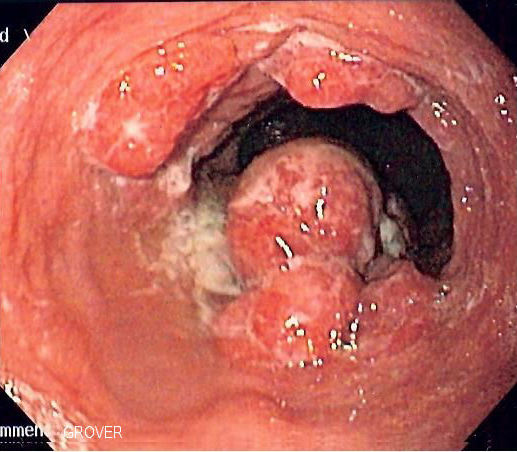
Esophageal cancer is a malignancy arising from the esophageal epithelium, comprising two principal histologic subtypes: (ESCC) and (EAC). The global burden is substantial: in 2022, at least 511,000 new cases were recorded worldwide, with 43,571 from Europe and 22,370 new cases in the United States in 2024 [1]A1c.
Mortality trends show a narrowing but persistent sex gap. Among European Union men, the esophageal cancer mortality rate declined from 5.34 to 4.99 per 100,000 between 2000-2004 and 2005-2009 (a 7% reduction), and is predicted to fall further to 4.5 per 100,000 by 2015 (approximately 22,300 deaths). For EU women, rates have remained low and stable, moving from 1.12 to 1.09 per 100,000, with a predicted 2015 rate of 1.1 per 100,000 (about 7400 deaths) [7]D5.
Geography and histology are tightly linked. In northern Europe, EAC is now the predominant histologic type among men, whereas ESCC remains more common in women and is still increasing in several countries [7]D5. Eastern populations have higher rates of squamous cell carcinoma, while adenocarcinoma predominates in the Western world; both subtypes show a male preponderance [1]A1c.
The disease typically afflicts older adults: the mean age across studies of cohorts ranges from 60.3 to 73.9 years [11]B2a. Age is a strong independent risk factor for postoperative pneumonia [11]B2a.
Several epidemiological patterns merit attention. Among individuals with a positive immunochemical fecal occult blood test (iFOBT) in a bowel cancer screening program, the incidence of esophageal cancer was 114.42 per 100,000, approximately 7.5 times higher than the background population rate of 36.08 per 100,000 [20]B3b. , though uncommon overall (pooled incidence 2.84%), occur significantly more often in adenocarcinoma (5.34%) than in squamous cell carcinoma (1.45%) [12]B2a. Among patients with metastatic disease at presentation, 24% have de-novo oligometastatic disease (≤5 metastases), and their median overall survival is 16 months (95% CI 13-21) [10]A1b.
These geographic and temporal patterns are driven by the distribution of key risk factors, which are discussed in the next section.
Pearl: A patient with a positive iFOBT has a 7.5-fold higher rate of esophageal cancer than the general population [20]B3b, consider in this group even when is negative.
Risk Factors and Prevention
- ▸Risk factors for esophageal cancer are histology-specific: metabolic syndrome and obesity drive EAC, while alcohol, smoking, and achalasia drive SCC.
- ▸Screening is effective in high-risk populations and is increasingly performed with minimally invasive tools such as the capsule-sponge device.
- ▸Modifiable risk factors (tobacco, alcohol, obesity, diet) are the primary prevention targets.
The incidence of esophageal cancer varies dramatically by geography and histology, reflecting distinct risk factor profiles for squamous cell carcinoma (SCC) and adenocarcinoma (EAC). The table below summarizes the key independent risk factors established in prospective studies.
| Risk Factor | HR/OR (95% CI) | Interpretation | Independent? | Reference |
|---|---|---|---|---|
| Metabolic syndrome (for EAC) | HR 1.24 (1.07-1.42) | 24% increased risk of EAC | Yes | [21]C4 |
| Obesity (for EAC) | HR 1.50 (1.24-1.82) | 50% increased risk of EAC | Yes | [21]C4 |
| Hyperglycemia (for EAC) | HR 1.14 (1.01-1.29) | 14% increased risk of EAC | Yes | [21]C4 |
| (ALD) | HR 2.18 (1.36-3.49) | 2.2-fold increased risk of EC | Yes | [27]B3b |
| MetALD (alcohol + metabolic) | HR 1.68 (1.17-2.42) | 68% increased risk of EC | Yes | [27]B3b |
| aHR 3.40 (1.25-9.22) | 3.4-fold increased risk of EC | Yes | [31]C4 | |
| adherence (for ESCC in men) | HR 0.57 per 2-point increment (0.41-0.80) | 43% reduced risk per 2-point increase in aMEDr score | Yes | [25]B2b |
| Smoking (for ESCC) | Significant (p = 0.044) | Risk factor | Yes | [32]B3b |
| High MCV (for ESCC) | Significant (p = 0.0018) | Risk factor | Yes | [32]B3b |
| Severe gastric atrophy (for ESCC) | Significant (p = 0.048) | Risk factor | Yes | [32]B3b |
| Positive iFOBT | Rate 114.42 per 100,000 vs 36.08 per 100,000 | 7.5 times higher risk of EC | Yes | [20]B3b |
| GLP-1 receptor agonist use | RR 0.46 (0.13-1.59) | Not associated with increased risk; may be protective | No (not significant) | [4]A1a |
Prevention
Prevention strategies target these modifiable risk factors. Tobacco cessation and alcohol avoidance remain the most powerful interventions for SCC, particularly in high-incidence regions like East Asia and sub-Saharan Africa. For EAC, weight reduction and management of metabolic syndrome are key. The Mediterranean diet, particularly adherence to the alternate Mediterranean diet score without alcohol (aMEDr), was associated with a 43% reduction in ESCC risk per 2-point increment in men [25]B2b. GLP-1 receptor agonists, increasingly used for diabetes and obesity, did not increase esophageal cancer risk (RR 0.46, 95% CI 0.13-1.59) [4]A1a; retrospective data suggest a possible protective effect, but this remains unconfirmed in randomized trials.
Screening
Screening for esophageal cancer is recommended in high-risk populations. The China guideline for the screening, early detection and early treatment of esophageal cancer (2022) provides evidence-based recommendations for screening population, technology, and procedure management [6]A1c. In Japan, screening endoscopy for ESCC in males over 50 with smoking and drinking history has shown increasing detection rates, with 95.2% of detected ESCCs at early stages [32]B3b. Minimally invasive screening tools, such as the capsule-sponge device paired with p53 immunohistochemistry, have shown high accuracy for ESCC (sensitivity 83.3%, specificity 95.4%) [24]C4. Public acceptance of screening is influenced by disease awareness, fear, belief in benefit, and practicalities [22]B2a. Notably, the rate of interval cancers after negative screening endoscopy is substantial (0.01% in a Korean cohort of 28 million), and endoscopist workload impacts detection rates: detection rates in the fourth quarter were half those in the first quarter [29]B3b.
These risk factors and screening strategies form the foundation for identifying high-risk individuals, which is further refined by genetic predisposition discussed in the next section.
Pearl: The sharp histologic dichotomy in risk factors means that screening strategies must be tailored: for SCC, target older men with smoking and alcohol history; for EAC, target those with obesity, metabolic syndrome, and GERD.
Genetics and Hereditary Predisposition
- ▸ALDH2 deficiency (rs671 GA/AA) synergizes with heavy alcohol consumption (≥30 g/d) to markedly increase esophageal cancer risk, but confers no increased risk in abstainers or light drinkers.
- ▸Androgen pathway SNPs (CYP17A1, JMJD1C) showed preliminary associations with BE and EAC but failed to replicate in independent samples.
- ▸Computational models predict many miRNA-disease associations for esophageal cancer, but these are not germline hereditary variants.
The risk conferred by established environmental exposures is modified by common genetic variants, most notably the ALDH2 Glu504Lys (rs671 G>A) polymorphism, which regulates acetaldehyde metabolism. This variant is almost exclusively present and highly prevalent among East Asians [35]C4. In carriers of the Lys/Lys and Glu/Lys genotypes, ALDH2 enzyme activity is reduced to nearly 0% and 17-38% of normal, respectively, leading to acetaldehyde accumulation after alcohol consumption [35]C4.
ALDH2 Polymorphism and Alcohol Consumption
In the China Kadoorie Biobank prospective cohort of 0.5 million adults aged 30-79 years, low-activity ALDH2 was not associated with increased esophageal cancer risk in the absence of alcohol consumption [35]C4. Among male weekly alcohol consumers, however, both self-reported flushing "soon after drinking" and the rs671 GA genotype were associated with significantly elevated risk. The adjusted hazard ratio for EC with rs671 GA versus GG was 3.31 (95% CI 1.94-5.67) [35]C4. The effect was confined to men consuming ≥30 g of alcohol per day; no increased risk was seen among light-to-moderate drinkers [35]C4. The HR for EC associated with each 15 g/d increment of alcohol was 1.28 (1.15-1.44) among men with a "soon" flushing response, compared with 1.12 (1.09-1.15) among those without (p_interaction = 0.047) [35]C4. For rs671 GA versus GG, the corresponding HRs were 1.41 (1.08-1.82) and 1.16 (1.06-1.27) (p_interaction = 0.493) [35]C4.
Self-reported facial flushing as a surrogate for inactive ALDH2 had low sensitivity (56.8%) but high specificity (88.4%) among male weekly alcohol consumers [35]C4. Adding past flushing response did not appreciably improve detection [35]C4. The population attributable fraction for EC that would be eliminated if men with rs671 GA who consumed ≥30 g/d changed to light-to-moderate consumption or abstinence was 7% [35]C4.
Androgen Pathway Gene Variants
Polymorphisms in genes within the androgen pathway have been investigated for Barrett's esophagus and esophageal adenocarcinoma. In a combined analysis of 14 studies (1,508 EAC, 2,383 BE, 2,170 controls), SNPs within CYP17A1 were associated with risk of BE in the sexes combined (p = 0.002) and in males (p = 0.003), but not in females separately [39]D5. SNPs within JMJD1C were associated with risk of EAC in females (p = 0.001) [39]D5. However, none of these associations replicated in a subsequent sample, and 14 other androgen-pathway genes showed no significant association after correction for multiple testing [39]D5.
Emerging miRNA Disease Associations
Computational models integrating miRNA functional similarity, disease semantic similarity, and known miRNA-disease associations have predicted numerous miRNAs linked to esophageal neoplasms. The HFHLMDA model achieved 90% confirmation of top 50 predictions, EGBMMDA confirmed 98% (49 of 50), and HLPMDA confirmed 47 of 50 candidate miRNAs in case studies [36]D5[37]D5[38]D5. These findings suggest a growing role for miRNAs as biomarkers, though they are not germline variants and are not yet incorporated into clinical hereditary risk assessment.
Pearl: The ALDH2 rs671 A allele increases esophageal cancer risk only in combination with alcohol consumption ≥30 g/day; self-reported flushing has low sensitivity (56.8%) for identifying this genotype, so genotyping should be considered for risk stratification in East Asian populations who drink heavily.
| Genotype / Flushing Response | HR (95% CI) | p_interaction |
|---|---|---|
| rs671 GA vs GG | 3.31 (1.94-5.67) | 0.376 |
| "Soon" flushing vs none | 1.45 (1.05-2.01) | 0.618 |
| Per 15 g/d alcohol, rs671 GA | 1.41 (1.08-1.82) | 0.493 |
| Per 15 g/d alcohol, "soon" flushing | 1.28 (1.15-1.44) | 0.047 |
Histopathology and Molecular Biology
- ▸Esophageal cancer has two main histologic subtypes, squamous cell carcinoma (ESCC) and adenocarcinoma (EAC), with distinct epidemiology, molecular drivers, and prognosis.
- ▸SCC histology is associated with higher recurrence risk and a more complex clinical course, often requiring careful selection for neoadjuvant therapy.
- ▸Molecular testing for HER2, PD-L1, and mismatch repair deficiency (dMMR) is essential for guiding systemic therapy, particularly in advanced EAC.
The genetic alterations described in the preceding section give rise to two main histologic subtypes, esophageal squamous cell carcinoma (ESCC) and esophageal adenocarcinoma (EAC), that differ fundamentally in epidemiology, molecular pathogenesis, and clinical behavior. Histologic subtype is the single most important determinant of prognosis and guides the choice of systemic therapy, with emerging molecular markers further refining treatment decisions.
Histologic Subtypes
Esophageal squamous cell carcinoma (ESCC) arises from the squamous epithelium lining the upper and middle esophagus. It predominates in Eastern populations and is strongly linked to tobacco and alcohol use [1]A1c. In Western cohorts, ESCC accounts for approximately 14% of cases [47]B3b. Esophageal adenocarcinoma (EAC) develops from Barrett metaplasia in the distal esophagus and gastroesophageal junction, is more common in Western countries, and constitutes 71-85% of cases in North American series [1]A1c[47]B3b[53]D5. Both subtypes show a male preponderance [1]A1c.
Rare histologies include liposarcoma, which most frequently presents as a well-differentiated tumor in the hypopharynx or cervical esophagus and carries a low risk of nodal or distant metastasis [44]B2a.
Molecular Drivers
Molecular profiling has identified distinct driver alterations in each subtype, though the evidence base for specific mutations is limited in the current literature. Key features include:
- ESCC: Frequent TP53 mutations, CDKN2A loss, and amplification of CCND1 or EGFR. These alterations drive cell cycle dysregulation and resistance to apoptosis.
- EAC: TP53 mutations are nearly universal, with frequent CDKN2A inactivation, HER2 amplification (in ~15-20%), and KRAS or SMAD4 mutations. HER2 overexpression defines a targetable subset.
- Mismatch repair deficiency (dMMR) is an emerging biomarker that may influence treatment choice, though definitive recommendations are not yet established [1]A1c.
- MicroRNA dysregulation has been implicated in malignant transformation: miR-374 and miR-574 are deregulated in esophageal mucosa after caustic injury and are linked to esophageal tumorigenesis [54]D5.
Prognostic and Therapeutic Implications
Histologic subtype independently affects prognosis and treatment selection:
- Recurrence risk: SCC histology is associated with a higher risk of recurrence after multimodality therapy (adjusted HR not reported in source) [51]B3b. Patients with SCC are also more likely to have surgery canceled after neoadjuvant treatment, reflecting a more complex oncologic and functional profile [49]C4.
- Therapy selection: For locally advanced disease, neoadjuvant chemoradiation (e.g., CROSS regimen) is effective for both subtypes, but the optimal neoadjuvant approach may differ. The ISDE guideline panel made no separate recommendation by histology due to insufficient evidence [1]A1c. For EAC, perioperative chemotherapy (e.g., FLOT) is often preferred, while SCC may be more radiosensitive.
- Molecular targets: HER2 testing is standard for advanced EAC to identify candidates for . PD-L1 expression guides immunotherapy use. dMMR status, though rare, predicts response to immune checkpoint inhibitors [1]A1c.
Table: Comparison of ESCC and EAC
| Feature | ESCC | EAC |
|---|---|---|
| Epidemiology | Higher in East Asia, Africa | Higher in Western countries |
| Risk factors | Tobacco, alcohol, caustic injury | GERD, obesity, |
| Typical location | Upper/middle esophagus | Distal esophagus, GEJ |
| Common mutations | TP53, CDKN2A, CCND1, EGFR | TP53, CDKN2A, HER2, KRAS, SMAD4 |
| Recurrence risk | Higher [51]B3b | Lower (relative) |
| Targetable markers | EGFR (rare), PD-L1 | HER2, PD-L1, dMMR |
Histologic subtype and molecular profile directly influence the pattern of metastasis and clinical behavior, SCC more often involves the airway and brain, while EAC preferentially spreads to liver and peritoneum. These distinctions will be further explored in the next section on clinical presentation.
Pearl: In patients with esophageal cancer, histologic subtype is not merely a pathologic label, it dictates the likelihood of nodal upstaging, the choice between neoadjuvant chemotherapy and chemoradiation, and the priority of molecular testing for HER2, PD-L1, and dMMR.
Clinical Presentation
- ▸Early-stage esophageal cancer is typically asymptomatic, with most patients presenting when the tumor is locally advanced or metastatic.
- ▸Progressive dysphagia (solids → liquids) is the cardinal symptom; weight loss, hoarseness, and cough indicate advanced disease.
- ▸Cachexia (low CXI) and sarcopenia are common signs that independently predict poor postoperative outcomes and survival.
The histologic subtypes described above, squamous cell carcinoma and adenocarcinoma, share a common challenge: the majority of early-stage tumors produce no symptoms, and the diagnosis is often made at an advanced stage [29]B3b. The clinical presentation therefore reflects the extent of local invasion, lymph node involvement, and distant metastasis.
Symptoms
- Dysphagia - progressive, first to solids then liquids; the most common presenting symptom.
- Odynophagia - pain on swallowing, often with advanced or ulcerated lesions.
- - occurs in >50% of patients at presentation.
- Regurgitation and vomiting of undigested food.
- Chest pain or retrosternal discomfort.
- Hoarseness - due to recurrent laryngeal nerve invasion.
- Cough, choking, or aspiration - may indicate tracheoesophageal fistula. Early-stage disease is typically asymptomatic, and lesions are frequently discovered incidentally during screening endoscopy [29]B3b.
Signs
- Cachexia - low cachexia index (CXI) and sarcopenia are common in advanced disease and independently predict poor survival [17]B3b[61]B2a.
- Anemia - from chronic occult blood loss.
- Supraclavicular lymphadenopathy (Virchow's node).
- Hepatomegaly or - from liver or peritoneal metastases.
- Pleural effusion - with metastatic spread.
- Positive fecal occult blood test - may be the first clue; in one series, esophageal cancer was found at a rate of 114.4 per 100,000 among iFOBT-positive patients, 7.5 times the general population rate [20]B3b.
Onset and Progression
Symptoms appear insidiously over weeks to months. Dysphagia initially to solids then progresses to liquids; weight loss accelerates as obstruction worsens. The median time from first symptom to diagnosis is often several months, contributing to the advanced stage at presentation.
Red Flags
- Complete dysphagia (inability to swallow saliva)
- Hematemesis or melena
- Severe malnutrition (e.g., >10% body weight loss in 6 months)
- Stridor or respiratory compromise (airway invasion)
Atypical Presentations
- - occur in 2.84% of patients (pooled incidence), with a higher rate in adenocarcinoma (5.34%) than squamous cell carcinoma (1.45%); may present with headache, seizures, or focal deficits [12]B2a.
- Positive iFOBT - up to 75% of patients with UGI cancer in one study had an unexplained positive fecal occult blood test, suggesting that a gastroscopy may be warranted in this setting [20]B3b.
Pearl: Esophageal cancer is most often asymptomatic in its early, curable stage; any new, progressive dysphagia in a patient over 50, especially with risk factors, should prompt immediate regardless of the absence of other alarm signs [29]B3b.
Biopsy and Histologic Diagnosis
- ▸Endoscopic biopsy has a 34-49% discrepancy rate with resection specimens, often underestimating disease severity.
- ▸Capsule-sponge sampling with cytology and p53 IHC offers a safe, high-accuracy screening alternative (sensitivity 83.3%, specificity 95.4%).
- ▸Immunohistochemical markers (MUC2, LI-cadherin, p53, CDKN2A) aid in histologic classification and risk stratification.
After clinical suspicion of esophageal cancer is raised, tissue acquisition for histologic confirmation is the critical next step. Endoscopic forceps biopsy remains the gold standard for initial diagnosis, but its accuracy is limited by sampling error and lesion heterogeneity. In superficial esophageal squamous neoplasms, the histopathologic discrepancy rate between forceps biopsy and endoscopic resection specimens is 34.5%, with upgrades (from HGIN to SCC or LGIN to SCC) accounting for 27 of 29 discordant lesions [72]C4. Similarly, in a large multicenter ESD registry, 48.7% of biopsies disagreed with the resection specimen, and 40.4% were underestimated; carcinoma was unsuspected on biopsy in 54.8% of cases [74]B3b. A larger lesion (≥22 mm) was a significant predictor of histologic upstaging [74]B3b.
Minimally Invasive Sampling Alternatives
The capsule-sponge device offers a safe, office-based alternative for detecting esophageal squamous neoplasia. In the EDEN trial, capsule-sponge sampling with cytologic assessment for intraepithelial neoplasia achieved accuracy 94.2% (95% CI 89.6-96.8%), sensitivity 83.3% (95% CI 60.8-94.2%), and specificity 95.4% [24]C4. Adding p53 immunohistochemistry to atypical cytology increased the odds ratio for neoplasia to 161.1 (95% CI 28.7-903.2) for a dual-positive test [24]C4. No adverse events occurred, and median acceptability was neutral-to-favorable [24]C4.
Adjunctive Optical Diagnostic Techniques
Optical coherence tomography (OCT) provides cross-sectional imaging of esophageal wall layers. In prospective studies, OCT identified intestinal metaplasia with sensitivity 81-97% and specificity 57-92%, and dysplasia/early cancer with sensitivity 68-83% and specificity 75-82% [70]A1a. For staging early squamous cell carcinoma, OCT accuracy exceeded 90% when compared to histologic resection specimens [70]A1a. However, current performance does not meet recommended thresholds to replace 4-quadrant biopsies in clinical practice [70]A1a.
Histopathologic and Immunohistochemical Assessment
Histologic classification of Barrett's-related neoplasia can be challenging. Immunohistochemical markers help distinguish between lesions: MUC2 and LI-cadherin expression differs significantly between normal glandular mucosa, Barrett's mucosa, intraepithelial neoplasia, and invasive carcinoma, but neither marker distinguishes low-grade from high-grade intraepithelial neoplasia [84]D5. Mutational profiling of biopsy tissue is increasingly feasible: CDKN2A/B and FHIT somatic copy number alterations are detected in 88% of progressors versus 24% of non-progressors, suggesting a role for early risk stratification [79]D5. In esophageal squamous cell carcinoma, MDM2 copy number increases occur in 9% of cases and are associated with poor prognosis [80]D5.
After neoadjuvant chemoradiotherapy, post-treatment biopsies have sensitivity of only 11.1% for detecting residual disease; an algorithm combining endoscopic category with biopsy results improves sensitivity to 64.8% and specificity to 95.9% for predicting pathological complete response [73]C4.
Pearl: When a biopsy shows low-grade or high-grade intraepithelial neoplasia, anticipate a 34-49% chance of histologic upgrade on endoscopic resection; a larger lesion (≥22 mm) and upper esophageal location are independent predictors of upstaging [72]C4[74]B3b.
| Technique | Target Condition | Sensitivity | Specificity | Accuracy |
|---|---|---|---|---|
| OCT for intestinal metaplasia | Barrett's esophagus | 81-97% | 57-92% | Not reported [70]A1a |
| OCT for dysplasia/early cancer | Esophageal neoplasia | 68-83% | 75-82% | Not reported [70]A1a |
| OCT for early SCC staging | T staging of SCC | Not reported | Not reported | >90% [70]A1a |
| Post-CRT biopsy alone | Residual disease after CRT | 11.1% | Not reported | Not reported [73]C4 |
| Post-CRT algorithm (endoscopic category + biopsy) | Pathologic CR prediction | 64.8% | 95.9% | 82.8% [73]C4 |
Imaging
- ▸PET/CT alters management intent in 13% of patients and reduces additional imaging by over 90% [95].
- ▸MRI with standardized T/V scores outperforms CT for detecting tracheal and vascular invasion (AUC 0.94-0.99 vs ≤0.71) [96].
- ▸DCE-MRI grading after neoadjuvant therapy identifies complete response with 100% PPV (grade 1) and improves accuracy when combined with PET/CT or endoscopy [97].
With histologic confirmation established, imaging defines the anatomic extent of disease and drives treatment decisions. The choice and sequencing of modalities follow guideline recommendations and the clinical question being asked.
Staging Modalities
CT of the chest and abdomen is the initial imaging study for locoregional and distant staging, though its accuracy for T stage is only 40-50% [1]A1c. CT-based T staging relies on indirect signs such as fat plane obliteration, organ deformity, and tumor contact angle; equivocal findings (cT3br) are common [96]C4.
EUS provides the best T and N staging among available modalities, but its accuracy is suboptimal. For esophageal adenocarcinoma, overall T staging accuracy is 65.5%; sensitivity and specificity vary by T category (Table). Nodal staging accuracy is 77.9%, with sensitivity 77.3% and specificity 67.4% [93]B2a. Despite these limitations, EUS remains essential for locoregional assessment, particularly when combined with fine-needle aspiration.
| T category | Sensitivity | Specificity | Accuracy |
|---|---|---|---|
| T1 | 64.7% | 89.1% | 89.6% |
| T2 | 35.7% | 89.2% | 87.1% |
| T3 | 82.5% | 83.0% | 87.0% |
| T4 | 38.6% | 94.0% | 66.4% |
| Data from meta-analysis of EUS staging in esophageal adenocarcinoma [93]B2a. |
PET/CT is recommended for detecting distant metastases and alters management in 13.2% of patients (major change in treatment intent), while reducing the need for additional imaging from 36.6% to 3.1% [95]B2b. Pretreatment metabolic parameters carry prognostic value: higher SUVmax is associated with worse PFS (HR 1.06, p=0.011) and RFS/DFS (HR 1.09, p=0.019); higher MTV with worse OS (HR 1.02, p=0.049); higher TLG with worse RFS/DFS (HR 2.02, p=0.022) [87]B2a. For cT2N0 disease, the ISDE guideline panel recommends both FDG-PET and EUS before treatment [1]A1c.
MRI offers superior soft-tissue contrast for assessing T4 invasion. A standardized MRI scoring system (T-score for tracheal invasion, V-score for vascular invasion) achieves AUCs of 0.94-0.99 for tracheal invasion and 0.88 for vascular invasion, compared to ≤0.71 for CT [96]C4. Inter-reader agreement is substantial to almost perfect (κ=0.77-1.00) vs moderate for CT (κ=0.37-0.49) [96]C4. MRI should be considered when CT findings are equivocal for T4 disease.
Restaging After Neoadjuvant Therapy
After neoadjuvant chemoradiotherapy, accurate restaging is critical to identify patients who may avoid surgery. DCE-MRI using a qualitative five-grade system shows high performance: grade 1 (compatible with complete response) yields 100% PPV and specificity; combining grades 1-2 increases sensitivity to 73.5% with specificity 88.9% and accuracy 78.8% [97]B3b. Adding DCE-MRI to PET/CT improves accuracy from 65.4% to 80.8% (p=0.03), and to endoscopy from 55.8% to 76.9% (p=0.005) [97]B3b. DWI sequences also predict response: responders show a significant percent increase in ADC during treatment (mean difference 21.06%, p<0.01) [88]B2a.
Surveillance for Recurrence
The pooled incidence of from esophageal carcinoma is 2.84% (95% CI 1.92-4.19%), with higher rates in adenocarcinoma (5.34%) vs squamous cell carcinoma (1.45%) [12]B2a. This supports regular brain imaging during follow-up, particularly for adenocarcinoma. CT-based radiomics models integrating clinical features predict early recurrence (within 1 year) after trimodal therapy, with AUC 0.809 in the validation cohort [92]A1b.
Emerging Techniques
Radiomics and deep learning applied to CT improve lymph node metastasis detection, with pooled AUC 87% in training sets [60]B2a. PET/CT radiomics combines PET and CT features for T stage, node, and pathological stage prediction, achieving AUCs of 0.815-0.837 in validation cohorts [99]B3b. A spectral CT-based nomogram incorporating neutrophil-to-lymphocyte ratio, clinical stage, effective atomic number, and normalized iodine concentration predicts neoadjuvant chemotherapy response with AUC 0.825 in the primary set [98]B3b. MRI-based radiomics models combining clinical factors predict DFS and OS with concordance indices of 0.714-0.730 [90]A1b.
Pearl: For suspected T4 disease, an MRI-based T/V score provides substantially better accuracy and inter-reader reliability than CT, and should be integrated into the staging algorithm when CT findings are equivocal [96]C4.
| T category | Sensitivity | Specificity | Accuracy |
|---|---|---|---|
| T1 | 64.7% | 89.1% | 89.6% |
| T2 | 35.7% | 89.2% | 87.1% |
| T3 | 82.5% | 83.0% | 87.0% |
| T4 | 38.6% | 94.0% | 66.4% |
| Data from meta-analysis of EUS staging in esophageal adenocarcinoma [93]B2a. |
Molecular Diagnostics and Biomarkers
- ▸HER2 testing is mandatory in metastatic gastroesophageal adenocarcinoma; the validated algorithm uses IHC with ISH confirmation for equivocal cases.
- ▸MSI-H is rare (0.5%-1.8%) but confers high TMB and predicts immunotherapy benefit; p16 overexpression in esophageal SCC is not HPV-driven.
- ▸Circulating biomarkers (ctDNA, miRNAs, supersulfides) show promise for early detection, prognosis, and treatment monitoring, with HomoCysSSH achieving AUC 0.93 for diagnosis.
Following imaging-based staging, molecular characterization of the tumor guides the selection of targeted and immunotherapeutic agents and provides prognostic information. The clinical utility of biomarkers in esophageal cancer depends on histologic subtype, stage, and the availability of matched therapies.
HER2 Testing
HER2 overexpression or amplification is the most established actionable biomarker in esophageal adenocarcinoma (EAC). In a nationwide Dutch cohort of 2846 patients with synchronous metastatic gastroesophageal adenocarcinoma treated with systemic therapy, HER2 testing increased from 18% in 2010 to 88% in 2016 [101]B3b. The overall HER2 positivity rate was 19% [101]B3b. Among HER2-positive patients, 77% received , and those treated with a trastuzumab-containing regimen had a median overall survival of 11.6 months versus 6.6 months with chemotherapy alone [101]B3b. The College of American Pathologists, American Society for Clinical Pathology, and American Society of Clinical Oncology recommend HER2 testing in all patients with metastatic gastroesophageal adenocarcinoma eligible for targeted therapy, using a validated algorithm: immunohistochemistry (IHC) 3+ is positive, 0 or 1+ is negative, and equivocal IHC 2+ requires confirmatory in situ hybridization (ISH) with a HER2:chromosome 17 ratio of ≥2 [101]B3b.
Microsatellite Instability and Genomic Biomarkers
Microsatellite instability-high (MSI-H) status is a rare but therapeutically relevant phenotype in esophageal carcinoma. In a multi-center real-world study of 1485 patients, the incidence of MSI-H was 0.49%-1.76% across centers [106]D5. All MSI-H tumors showed loss of PMS2 expression, with concurrent MLH1 loss in 75%; whole-exome sequencing revealed high tumor mutational burden (19.12-32.68 mutations/Mb) [106]D5. MSI-H is a biomarker for immune checkpoint inhibitor response, irrespective of histology. Other genomic alterations with prognostic significance include:
- MDM2 copy number increase - detected in 9% of ESCC cases, associated with shorter overall survival (p = 0.033) [80]D5.
- CDKN2A/B and FHIT deletions - found in 88% of nondysplastic Barrett's esophagus tissues from patients who progressed to dysplasia/adenocarcinoma, versus 24% in nonprogressors (p = 0.0004), suggesting a role as early progression biomarkers [79]D5.
- p16 overexpression - present in 9% of high-grade esophageal squamous cell carcinomas and 100% of small-cell carcinomas, but driven by RB1 pathway inactivation rather than HPV infection, and not predictive of favorable outcome [112]D5.
Circulating Biomarkers
Liquid biopsies offer a non-invasive approach to diagnosis, prognosis, and treatment monitoring.
Circulating tumor DNA (ctDNA). In 86 patients with previously treated metastatic gastroesophageal adenocarcinoma, the highest baseline ctDNA levels (top 20%) were associated with significantly shorter progression-free survival (median 1.8 vs. 4.6 months; HR 2.2, p = 0.005) and overall survival (median 5.5 vs. 9.9 months; HR 2.2, p = 0.004) [100]B2b. In resectable EAC (stage II/III), combining amplicon sequencing and shallow whole-genome copy number analysis detected ctDNA in 50.5% of patients at baseline; non-clearance of a baseline variant or ichorCNA >3% in pre-surgery samples was associated with a high risk of early progression (HR 4.58, 95% CI 2.22-9.46, p < 0.001) [107]D5.
MicroRNAs (miRNAs). Plasma miR-21 is significantly elevated in ESCC patients, while miR-31 and miR-375 are decreased. For differentiating ESCC from healthy controls, miR-31 and miR-375 each showed 87.5% sensitivity and 98.4%-100% specificity [103]B3b. Serum miR-18a and miR-21 have area under the curve (AUC) values of 0.948 and 0.914, respectively, outperforming conventional tumor markers [114]B3b.
Circulating tumor cells (CTCs). CTCs are detected in 84.6% of ESCC patients; TWIST-positive CTCs (75% of patients) are associated with poor histologic differentiation [108]D5.
Emerging Biomarkers
- Supersulfides. In a study of 50 esophageal cancer patients and 30 controls, plasma homocysteine hydropersulfide (HomoCysSSH) distinguished cancer from healthy subjects with AUC 0.93, sensitivity 89%, specificity 96% [102]B3b. Cysteine hydropersulfide (CysSSH) in exhaled breath condensate (EBC) had AUC 0.71, offering a non-invasive diagnostic alternative [102]B3b.
- Oncostatin M (OSM). OSM expression is elevated in ESCA tissues and correlates with advanced stage, lymph node metastasis, and poor prognosis; in vitro knockdown reduces proliferation and migration [109]D5.
- Stemness gene signatures. A prognostic model incorporating stemness genes, with RBBP7 as the most influential factor, accurately predicts 1-, 2-, and 3-year survival, outperforming staging [113]D5.
Integration with Staging
Molecular biomarkers complement anatomic staging by identifying patients most likely to benefit from targeted therapy (HER2, MSI-H) or immunotherapy (MSI-H, high TMB), and by providing early readouts of treatment response or resistance (ctDNA). The choice of which biomarkers to test is guided by histology, stage, and available therapeutic options, and results should be incorporated into the multidisciplinary treatment planning discussed in the next section.
Pearl: In metastatic gastroesophageal adenocarcinoma, HER2 testing is mandatory, the nationwide increase from 18% to 88% over 6 years paralleled a survival improvement from 6.9 to 7.9 months, highlighting the impact of biomarker-guided therapy [101]B3b.
| Biomarker | Specimen | Clinical Utility | Key Finding | Reference |
|---|---|---|---|---|
| HER2 (IHC/ISH) | Tumor tissue | Predicts trastuzumab benefit in metastatic EAC | Positivity rate 19%; OS 11.6 vs 6.6 mo with trastuzumab | [101]B3b |
| MSI-H | Tumor tissue | Predicts immunotherapy response | Incidence 0.5-1.8%; all MSI-H tumors show PMS2 loss | [106]D5 |
| ctDNA (methylation ddPCR) | Plasma | Prognosis, early treatment monitoring | High baseline ctDNA: PFS 1.8 vs 4.6 mo, OS 5.5 vs 9.9 mo | [100]B2b |
| ctDNA (amplicon + sWGS) | Plasma | Prognosis in resectable EAC | Non-clearance pre-surgery: HR 4.58 for early progression | [107]D5 |
| miR-31, miR-375 | Plasma | Diagnosis of ESCC | Sensitivity 87.5%, specificity 98.4-100% | [103]B3b |
| HomoCysSSH | Plasma | Diagnosis | AUC 0.93, sensitivity 89%, specificity 96% | [102]B3b |
| CysSSH | Exhaled breath condensate | Non-invasive diagnosis | AUC 0.71 | [102]B3b |
| MDM2 copy number increase | Tumor tissue | Prognosis | Detected in 9% ESCC; associated with worse OS | [80]D5 |
| CDKN2A/B + FHIT deletions | BE tissue | Risk of progression to DAC | 88% in progressors vs 24% in nonprogressors | [79]D5 |
Staging
- ▸Accurate staging integrates CT, EUS, PET/CT, and increasingly MRI, but all modalities have limitations, particularly for nodal assessment; occult LNM occurs in up to 48% of cT2N0 patients.
- ▸Risk stratification using tumor differentiation, size, SUVmax, and LVI helps identify patients who may safely undergo primary surgery versus those who benefit from neoadjuvant therapy.
- ▸Pathologic complete response after neoadjuvant therapy offers the best prognosis (5-year OS 75.1%), but residual nodal disease substantially worsens survival (ypT+N+ 5-year OS 26.1%).
The molecular and histologic characterization of the tumor must be integrated with anatomic staging to guide treatment decisions. Accurate staging is the cornerstone of esophageal cancer management, determining whether a patient is a candidate for endoscopic resection, neoadjuvant therapy, or palliative treatment, and every staging modality has limitations that clinicians must understand.
Classification and Stage Groups
Staging follows the 8th edition, which incorporates histologic type and grade into the stage grouping. For example, T2N0M0 adenocarcinoma is upstaged from Stage IC to IIA in the presence of poor differentiation, and squamous cell cancer is upstaged from Stage IB to IIA unless the cells are well-differentiated [1]A1c. After neoadjuvant therapy, the ypStage (pathologic stage after therapy) is used, but it may not fully capture prognosis: patients with ypT0N0 (pathologic complete response, pCR) have a 5-year overall survival (OS) of 75.1%, whereas those with ypT+N0 have 42.4%, ypT0N+ 54.9%, and ypT+N+ 26.1% [120]B3b. Even within ypStage I, survival differs between ypT0N0 and ypTis-2N0 [120]B3b.
Staging Modalities and Accuracy
No single modality provides perfect staging, and the accuracy of clinical staging (cTNM) compared with pathologic staging (pTNM) is often poor. CT has an accuracy of only 40-50% for T stage [1]A1c. Endoscopic ultrasound (EUS) performs better but still under 90% for nodal status, and occult nodal metastasis rates of up to 50% have been reported [1]A1c. Low-dose hydro-CT with iterative reconstruction offers a sensitivity of 93%, specificity of 100%, positive predictive value (PPV) of 100%, and negative predictive value (NPV) of 88% for esophageal cancer diagnosis, with near-perfect inter-reader agreement for T, N, and M stages (κ > 0.90) [64]B3b.
PET/CT has a significant impact on management: in a prospective registry, major changes (e.g., curative to palliative or vice versa) occurred in 13.2% of patients, and minor changes in 24.1% [95]B2b. Curative-intent therapy based on PET/CT was associated with a median OS of 3.5 years versus 0.9 years for palliative intent [95]B2b.
MRI outperforms CT for detecting tracheal and vascular invasion in suspected T4 disease. The area under the ROC curve (AUC) for tracheal invasion was 0.943-0.990 for MRI versus 0.529-0.706 for CT; for vascular invasion, MRI AUC was 0.878 versus ≤0.706 for CT [96]C4. Inter-reader agreement was substantial to almost perfect for MRI (κ = 0.771-1.000) but only moderate for CT (κ = 0.369-0.487) [96]C4. A combined MRI score (T-score or V-score) achieved specificity ≥ 0.88 without loss of sensitivity, supporting its use when CT findings are equivocal [96]C4.
Optical coherence tomography (OCT) may identify intestinal metaplasia and dysplasia, but its accuracy (sensitivity 68-83%, specificity 75-82% for dysplasia and early cancer) may not meet thresholds to replace four-quadrant biopsies [70]A1a. OCT may be more accurate than EUS for staging early esophageal cancer, but randomized trials are lacking [70]A1a.
The Challenge of Nodal Staging
Occult lymph node metastasis (LNM) is the most critical staging pitfall. In patients with clinical T1-T2 N0 esophageal squamous cell carcinoma (ESCC) undergoing upfront surgery, occult LNM was found in 21.8% [91]A1b. A risk model identified four independent predictors: tumor maximum standardized uptake value (SUVmax) > 3.8, higher histological differentiation grade (poorer differentiation), tumor length > 25 mm, and advanced clinical T stage [91]A1b. The risk scoring system showed high accuracy (AUC 0.81 in the training set, 0.743 in the testing set) [91]A1b.
For cT2N0 disease, the correlation between clinical and pathologic staging is poor. Accurate staging was reported in only 6-42.8% of cases, and upstaging after histopathologic review occurred in 21.4-63.4%, with nodal upstaging in up to 48% of patients [1]A1c. In early esophageal adenocarcinoma, the overall metastatic rate (including LNM and distant metastases) increases with depth: sm1 11.9%, sm2 27.3%, sm3 32.1% [124]C4. Lymph node metastases were absent in m1 and m2 tumors and rare in m3 (1/18) and m4 (5/21), but more frequent in sm2 (11/44) and sm3 (18/78) [124]C4.
Risk Stratification for Occult Disease and Treatment Selection
Given the high rate of understaging, the ISDE guideline panel recommends neoadjuvant therapy followed by surgery for most patients with cT2N0 esophageal cancer (conditional recommendation, low certainty of evidence) [1]A1c. The panel identified low-risk features that may allow primary surgery alone: small (< 2 cm), well-differentiated tumor, no lymphovascular invasion (LVI), and high confidence in staging workup [1]A1c. High-risk features favoring neoadjuvant therapy include poor differentiation, LVI, tumor length ≥ 2 cm, and higher SUVmax [1]A1c[91]A1b.
Staging After Neoadjuvant Therapy
Pathologic complete response (pCR) after neoadjuvant therapy offers the best prognosis, with a 5-year OS of 75.1% [120]B3b. However, residual nodal disease substantially worsens survival: patients with ypT0N+ (pCR of primary but residual nodal metastases) had a 5-year OS of 54.9%, and those with ypT+N+ had only 26.1% [120]B3b. The presence of a positive supraclavicular lymph node (cSCN+ or pSCN+) did not independently predict OS after three-field lymph node dissection in ESCC [121]B3b.
Clinical Implications
Staging accuracy directly drives treatment strategy. Understaging leads to undertreatment and higher recurrence risk, while overstaging exposes patients to unnecessary neoadjuvant therapy. MRI should be integrated when CT findings are equivocal for T4 disease [96]C4. PET/CT frequently changes management and should be part of the staging workup [95]B2b. The management section that follows builds on these staging principles to outline the multimodal treatment algorithm.
Pearl: The critical challenge in esophageal cancer staging is the high rate of occult nodal metastasis (up to 48% in cT2N0), which makes neoadjuvant therapy the preferred approach for most patients unless low-risk features, small size, well-differentiated histology, no LVI, and high-confidence staging, are all present [1]A1c.
| Modality | Finding | Sensitivity | Specificity | PPV | NPV | Agreement |
|---|---|---|---|---|---|---|
| Hydro-CT | Diagnosis of EC | 93% [64]B3b | 100% [64]B3b | 100% [64]B3b | 88% [64]B3b | κ > 0.90 [64]B3b |
| MRI (T-score) | Tracheal invasion | - | - | - | - | AUC 0.943-0.990 [96]C4 |
| CT (T-stage) | Tracheal invasion | - | - | - | - | AUC 0.529-0.706 [96]C4 |
| MRI (V-score) | Vascular invasion | - | - | - | - | AUC 0.878 [96]C4 |
| CT (V-stage) | Vascular invasion | - | - | - | - | AUC ≤ 0.706 [96]C4 |
| OCT | Dysplasia and early cancer | 68-83% [70]A1a | 75-82% [70]A1a | - | - | - |
| Risk Factor | Threshold | Odds/Hazard | Source |
|---|---|---|---|
| SUVmax | > 3.8 | Independent predictor (P = 0.002) | [91]A1b |
| Tumor length | > 25 mm | Independent predictor (P < 0.001) | [91]A1b |
| Clinical T stage | Advanced | Independent predictor (P < 0.001) | [91]A1b |
| Lymphovascular invasion | Present | High-risk feature (expert consensus) | [1]A1c |
| Tumor size (cT2N0) | ≥ 2 cm | High-risk feature (expert consensus) | [1]A1c |
Management Overview
- ▸Treatment selection depends on stage, histology, and patient fitness, with endoscopic therapy for early disease, neoadjuvant therapy for resectable disease, definitive chemoradiotherapy for unresectable tumors, and systemic therapy (with or without immunotherapy) for advanced disease.
- ▸Pembrolizumab plus cisplatin/5-FU improves OS in advanced esophageal cancer (HR 0.65, 60-month OS 24.0% vs 8.5%) but is not cost-effective at current pricing in Japan.
- ▸Conversion surgery after effective induction therapy significantly improves survival in initially unresectable esophageal cancer (1-year OS RR 1.53, 3-year RR 2.03).
Staging, histology, and patient fitness jointly determine the treatment strategy for esophageal cancer. The paradigm is multimodal: endoscopic resection for early disease, neoadjuvant or perioperative therapy for locoregionally advanced disease, definitive chemoradiotherapy for unresectable tumors, and systemic therapy, increasingly including immunotherapy, for metastatic or recurrent cancer. Dedicated sections on each modality are linked below; this overview provides the framework for selecting among them.
Treatment Algorithm by Stage and Histology
The initial decision is whether the tumor is amenable to curative-intent treatment. The table below summarizes the standard approach by stage grouping.
| Stage grouping | Preferred approach | Key considerations |
|---|---|---|
| T1a (high-grade dysplasia, intramucosal cancer) | Endoscopic eradication therapy (EET) | EET is strongly recommended for high-grade dysplasia and intramucinal adenocarcinoma [2]A1c. For low-grade dysplasia, a conditional recommendation supports EET, but surveillance is an option for patients who value avoiding harms [2]A1c. |
| T1b (submucosal invasion) | EET or | Depends on depth of invasion, lymph node risk; for visible lesions, focal EMR plus ablation is preferred over stepwise EMR [2]A1c. |
| cT2N0M0 | Neoadjuvant therapy followed by surgery (conditional recommendation) | The ISDE guideline gives a conditional recommendation for neoadjuvant therapy over primary surgical resection in this group [1]A1c. |
| T3-4a or N+ (resectable) | Neoadjuvant chemoradiotherapy (nCRT) or perioperative chemotherapy | nCRT improves R0 resection rate and pathologic complete response (pCR) rate compared with neoadjuvant chemotherapy alone, but long-term survival benefit is not clearly different [126]A1a. |
| Unresectable locally advanced (T4b, bulky N+) | Definitive chemoradiotherapy (dCRT) | Median OS is approximately 25 months; event-free survival is a strong surrogate for OS (R² = 0.80) [129]A1a. |
| Metastatic (M1) | Systemic therapy; + chemotherapy for PD-L1 CPS ≥10 | The Japanese subgroup of KEYNOTE-590 showed median OS 17.7 vs 11.7 months (HR 0.65) with pembrolizumab plus / ; 60-month OS rate 24.0% vs 8.5% [127]A1b. |
Neoadjuvant and Perioperative Therapy
For resectable esophageal cancer, preoperative therapy improves survival compared with surgery alone. The choice between nCRT and neoadjuvant chemotherapy (nCT) is debated. A meta-analysis of three randomized trials found that nCRT significantly increases R0 resection rate and pCR rate, but 3- and 5-year overall survival did not differ between the approaches [126]A1a. Thus, the addition of radiotherapy to chemotherapy does not clearly confer a survival advantage, despite improving local response. In patients with cT2N0 disease, the ISDE guideline makes a conditional recommendation for neoadjuvant therapy followed by surgery over primary surgical resection, acknowledging that staging uncertainty is high [1]A1c.
Definitive Chemoradiotherapy for Unresectable Disease
For patients with locally advanced, unresectable tumors (T4b, bulky nodal disease, or medically inoperable), dCRT is the standard of care. A correlation analysis of 11 randomized trials (N = 2812) demonstrated that event-free survival (EFS) is a strong surrogate for overall survival (R² = 0.80; slope 0.91; 95% CI 0.62-1.20) [129]A1a. Median EFS was 17.5 months and median OS was 25.1 months in these trials, supporting the use of EFS as an endpoint in future dCRT studies. Similarly, for resectable disease, recurrence-free survival or disease-free survival shows a strong correlation with OS (ρ = 0.89) [138]B2a, suggesting that these endpoints can accelerate trial completion.
Immunotherapy in Advanced Disease
First-line pembrolizumab plus chemotherapy has become a standard for advanced esophageal cancer, particularly in patients with PD-L1 combined positive score (CPS) ≥10. In the Japanese subgroup of KEYNOTE-590, with median follow-up of 60.6 months, the combination improved median OS from 11.7 to 17.7 months (HR 0.65; 95% CI 0.45-0.94) and raised the 60-month OS rate from 8.5% to 24.0% [127]A1b. Objective response rate was 56.8% vs 38.8%. However, cost-effectiveness analyses from Japan show an ICER of $176,479 per QALY in the overall population and $126,862 in the CPS ≥10 subgroup, exceeding the willingness-to-pay threshold of $50,000-$100,000, so the addition is not cost-effective in that setting [136]A1b.
Special Populations and Conversion Surgery
Older adults (≥70 years): In a target trial emulation of 432 patients with ESCC, concurrent chemoradiotherapy (CCRT) was associated with lower treatment completion rates (adjusted RR 0.86) and higher grade ≥3 toxicity (38.1% vs 17.8%) compared with radiotherapy alone [133]A1b. Effects were more pronounced in those aged ≥75, with Charlson Comorbidity Index ≥6, or frail (G8 ≤14). These data support careful patient selection and shared decision-making when considering intensification.
Initially unresectable disease: After effective conversion therapy, conversion surgery improves survival compared with continued non-surgical treatment. A meta-analysis of 10 studies (973 patients) found that conversion surgery was associated with significantly better OS at 1 year (RR 1.53), 2 years (RR 1.65), and 3 years (RR 2.03) [132]B2a. Postoperative complications included respiratory issues, anastomotic leakage, and recurrent laryngeal nerve injury.
Prehabilitation and Supportive Care
Multimodal prehabilitation, combining exercise, nutrition, and psychosocial support, during neoadjuvant chemotherapy is feasible and maintains cardiorespiratory fitness and functional capacity [9]C4. A 12-week walking and dietary education program improved rest-activity circadian rhythm and sleep quality at 6 months [137]A1b.
Thromboprophylaxis: Prolonged (1-month) low-molecular-weight after esophagectomy did not reduce thromboembolic events compared with standard in-hospital prophylaxis in a randomized trial. However, preoperative prothrombin fragment 1+2 (F1+2) levels predicted postoperative VTE (OR 1.64 per 50 pmol/L increase) [8]A1b, suggesting a role for risk stratification.
occurs after esophagectomy in a pooled prevalence of 27% (95% CI 14-39%), but rises to 67% when assessed by specialized questionnaires [135]A1a. Awareness and dietary counseling are important to mitigate its impact on quality of life.
Controversies and Guideline Disagreement
| Question | Position A | Position B | Strength | Implication |
|---|---|---|---|---|
| Neoadjuvant CRT vs CT for resectable EC | CRT improves pCR and R0 resection but not survival [126]A1a | CT alone may be preferred if toxicity is a concern | No consensus; both are NCCN options | Shared decision-making based on histology, performance status, and institutional preference |
| EET for low-grade dysplasia | Conditional recommendation in favor of EET [2]A1c | Surveillance is reasonable for patients who prioritize avoiding harms [2]A1c | Conditional recommendation | Patient preference is central |
| Cost-effectiveness of pembrolizumab + chemo | ICER exceeds WTP in Japan [136]A1b | Accepted in US and UK guidelines | Not cost-effective in Japan | May affect access; biomarker-based selection (CPS ≥10) improves ICER |
Pearl: For resectable esophageal cancer, neoadjuvant chemoradiotherapy improves pathologic response and R0 resection but does not clearly improve overall survival compared with neoadjuvant chemotherapy alone; in older or frail patients, radiotherapy alone may be better tolerated than concurrent chemoradiotherapy.
Management by Modality — Dedicated Pages
Detailed, modality-specific management is covered on dedicated pages:
- Esophageal Cancer Surgical Management — operations by stage, approach, resectability, adjuvant triggers
- Esophageal Cancer Radiation Management — EBRT, brachytherapy, concurrent chemoradiation, dose/fractionation, OAR constraints
- Esophageal Cancer Systemic Therapy — perioperative / definitive chemotherapy, targeted therapy, immune checkpoint inhibitors
- Esophageal Cancer Palliative Care — symptom management, stenting/nutrition, early integration, end-of-life
- Esophageal Cancer Surveillance and Follow-up — post-treatment surveillance schedule, late toxicity, survivorship
- Esophageal Cancer Recurrent and Metastatic Disease — locoregional salvage, systemic therapy for advanced disease, oligometastatic
| Stage grouping | Preferred approach | Key considerations |
|---|---|---|
| T1a (HGD, intramucosal) | Endoscopic eradication therapy (EET) | Strong recommendation for HGD; conditional for LGD [2]A1c |
| T1b (submucosal) | EET or esophagectomy | Focal EMR + ablation preferred over stepwise EMR [2]A1c |
| cT2N0M0 | Neoadjuvant therapy + surgery | Conditional recommendation over primary surgery [1]A1c |
| T3-4a or N+ (resectable) | nCRT or perioperative chemotherapy | nCRT improves pCR and R0 but not OS [126]A1a |
| Unresectable locally advanced | Definitive CRT | Median OS ~25 months; EFS is surrogate for OS [129]A1a |
| Metastatic | Systemic therapy ± pembrolizumab | Pembrolizumab + chemo for CPS ≥10 [127]A1b |
| Question | Position A | Position B | Strength | Implication |
|---|---|---|---|---|
| nCRT vs nCT for resectable EC | CRT improves pCR and R0 but not OS [126]A1a | CT alone may be preferred if toxicity is a concern | No consensus | Shared decision-making |
| EET for low-grade dysplasia | Conditional recommendation for EET [2]A1c | Surveillance is reasonable | Conditional recommendation | Patient preference central |
| Cost-effectiveness of pembrolizumab + chemo | ICER > WTP in Japan [136]A1b | Accepted in US/UK | Not cost-effective in Japan | Biomarker selection improves ICER |
History and Evolution of Treatment
- ▸Neoadjuvant therapy followed by surgery is the standard for locally advanced esophageal cancer, supported by ISDE guidelines [1].
- ▸Immunotherapy (pembrolizumab plus chemotherapy) provides durable survival benefit in advanced disease, with a 5-year OS rate of 24.0% vs 8.5% (NNT=7) [127].
- ▸Endoscopic techniques (ESD, biodegradable stents) and supportive care (prehabilitation, exercise, famotidine) have improved outcomes and quality of life, but careful patient selection is needed for older adults [133].
Building on the multimodal framework outlined above, the current standard of care for esophageal cancer has emerged through several pivotal shifts in surgical technique, systemic therapy, and endoscopic management, each grounded in landmark trials that redefined practice.
Surgery and Lymphadenectomy
The extent of lymphadenectomy has long been debated. Historically, 2-field lymphadenectomy (2FL) included mediastinal and abdominal nodes, while 3-field lymphadenectomy (3FL) added bilateral supraclavicular dissection. However, inconsistent definitions have complicated comparisons [143]B2b. Recent evidence, including meta-analyses and large multicenter retrospective studies, has not demonstrated a clear survival benefit of prophylactic 3FL over contemporary 2FL [143]B2b. The ongoing randomized phase III JCOG2013 trial is expected to clarify the therapeutic value of prophylactic supraclavicular dissection [143]B2b. Current Japanese guidelines weakly recommend 3FL for upper and middle thoracic esophageal cancer [143]B2b.
Systemic Therapy: From Chemotherapy to Immunotherapy
Early meta-analyses of -based combination chemotherapy showed no significant survival benefit when used as adjuvant or neoadjuvant therapy (odds ratio 0.96, 95% CI 0.75-1.22) [139]A1a. This led to the adoption of neoadjuvant chemoradiation as the standard for locally advanced disease. The International Society for Diseases of the Esophagus (ISDE) now conditionally recommends neoadjuvant therapy followed by surgery over primary surgical resection for cT2N0M0 esophageal cancer [1]A1c.
The landmark KEYNOTE-590 trial established immunotherapy as a cornerstone for advanced disease. In the Japanese subgroup, first-line 200 mg every 3 weeks plus chemotherapy (cisplatin 80 mg/m² and 800 mg/m²/day) significantly improved overall survival compared with placebo plus chemotherapy (median OS 17.7 vs 11.7 months; HR 0.65, 95% CI 0.45-0.94) [127]A1b. The 60-month OS rates were 24.0% and 8.5%, respectively, yielding an NNT of 7 to prevent one death over 5 years [127]A1b. Progression-free survival also improved (HR 0.57, 95% CI 0.39-0.83), with 60-month PFS rates of 16.9% vs 0% [127]A1b.
Endoscopic and Minimally Invasive Approaches
Endoscopic submucosal dissection (ESD) has become the preferred treatment for early esophageal neoplasms. A post hoc analysis of a multicenter RCT comparing insulated-tip knife (ESD-IT) and needle-type knife (ESD-N) found comparable procedural times (51.0 vs 49.5 minutes), complete resection rates (90.5% vs 90.7%), and complication rates (1.8% vs 3.7%) [145]A1b. However, the operator handover rate due to difficulty was significantly higher with ESD-N (13.0% vs 0.6%, P=0.001), which improved with traction assistance [145]A1b.
For stricture prevention after extensive ESD, a randomized trial compared intralesional triamcinolone 100 mg injection with oral prednisolone (30 mg/day tapered over 8 weeks). The oral group required significantly fewer dilation sessions (5 vs 19, P=0.04), though stricture rates were similar (66.7% vs 40%, P=0.27) [128]A1b. For refractory benign , a biodegradable stent achieved dysphagia improvement maintained at 3 months in 46.7% (95% CI 28.3-65.7) of patients, with median dysphagia-free survival of 98 days [141]B2b. However, caution is warranted in patients with prior chemoradiotherapy, as one fatal esophago-left atrium fistula occurred [141]B2b.
Supportive Care and Rehabilitation
Multimodal prehabilitation (exercise, nutrition, psychosocial support) during neoadjuvant chemotherapy is feasible, with compliance rates of 72% for supervised and 77% for home-based exercise, and both groups maintained cardiorespiratory fitness [9]C4. A nationwide Swedish RCT showed that a 12-week home-based exercise program 1 year after surgery significantly improved lower extremity strength (mean difference 4.48 vs 2.73, P=0.03) [140]A1b. A 12-week walking and dietary education program improved rest/activity circadian rhythm and total sleep time at 3 months (Cohen's d=0.86) [137]A1b.
Weight loss during neoadjuvant chemotherapy is an independent risk factor for postoperative infectious complications: >5% weight loss increased the odds ratio to 2.69 (95% CI 1.12-6.46, P=0.027) [15]B2b. Famotidine 40 mg orally daily before radiotherapy sessions reduced thrombocytopenia (P<0.0001) and preserved lymphocyte and platelet counts [144]A1b. A randomized trial found no benefit of prolonged (1-month) thromboprophylaxis after over standard in-hospital prophylaxis, though preoperative prothrombin fragment 1+2 levels predicted venous thromboembolism (odds ratio 1.64 per 50 pmol/L increase, 95% CI 1.17-2.54) [8]A1b. Non-placement of a cervical drainage tube after McKeown esophagectomy did not demonstrate non-inferiority to placement for anastomotic leakage (risk difference -0.100, P=0.0591) [142]A1b.
Special Populations: Older Adults
In patients aged ≥70 years with esophageal squamous cell carcinoma, concurrent chemoradiotherapy (CCRT) was associated with significantly lower treatment completion rates (adjusted risk ratio 0.86, 95% CI 0.79-0.93) and higher grade ≥3 toxicity (38.1% vs 17.8%; aRR 2.01) compared with radiotherapy alone [133]A1b. Effects were more pronounced in those aged ≥75 years, with Charlson Comorbidity Index ≥6, or frail (G8 ≤14) [133]A1b. These findings highlight the need for careful patient selection.
Controversies and Guideline Disagreement
| Question | Position A | Position B | Strength | Implication |
|---|---|---|---|---|
| Prophylactic 3FL vs 2FL for thoracic esophageal cancer | Japanese guidelines weakly recommend 3FL for upper/middle tumors [143]B2b | Recent evidence shows no clear survival benefit of 3FL over contemporary 2FL [143]B2b | Conditional | Await JCOG2013 trial results [143]B2b |
| Definitive RT vs CCRT for older adults (≥70 years) | CCRT offers numerically higher short-term tumor response [133]A1b | RT alone has higher completion rates and lower toxicity (grade ≥3: 17.8% vs 38.1%) [133]A1b | Strong for RT in frail patients | Shared decision-making essential [133]A1b |
These treatment advances have directly influenced prognostic outcomes, as discussed in the next section.
Pearl: Endoscopic techniques (ESD, biodegradable stents) and supportive care (prehabilitation, exercise, famotidine) have improved outcomes and quality of life, but careful patient selection is needed for older adults [133]A1b.
Prognosis and Prognostic Factors
- ▸Overall 5-year survival remains <20% globally, but reaches 92% after ESD for early-stage disease and 24% with pembrolizumab plus chemotherapy for advanced disease.
- ▸Beyond TNM stage, validated prognostic factors include cachexia index (CXI), weight loss >5% during neoadjuvant therapy, PET/CT metabolic parameters (SUVmax, MTV, TLG), and occurrence of immune-related adverse events.
- ▸RFS/DFS and EFS are strongly correlated with OS and serve as valid surrogate endpoints in clinical trials for resectable and unresectable disease, respectively.
Despite advances in multimodality therapy, the overall 5-year survival for esophageal cancer remains below 20% globally [135]A1a. Outcomes vary substantially by stage at diagnosis, histologic subtype, and treatment response. For early-stage disease managed with endoscopic submucosal dissection (ESD), 5-year disease-free survival (DFS) reaches 91.7% [119]B3b. In locally advanced disease, neoadjuvant therapy followed by surgery yields 5-year overall survival (OS) rates approaching 45-50% [146]B2b. For advanced or metastatic disease, first-line plus chemotherapy achieved a 5-year OS of 24.0% compared with 8.5% for chemotherapy alone (HR 0.65, 95% CI 0.45-0.94) in the Japanese KEYNOTE-590 subgroup [127]A1b.
Survival by Stage and Treatment
Survival varies markedly by treatment strategy, particularly in patients with cervical lymph node metastasis (CLNM). In a nationwide Dutch cohort, median OS was 24 months for neoadjuvant therapy followed by surgery, 18 months for definitive chemoradiotherapy (dCRT), 15 months for chemotherapy alone, 7 months for palliative radiotherapy, and 3 months for best supportive care. The 3-year OS rates were 38%, 21%, 10%, 2%, and 1%, respectively. Neoadjuvant therapy followed by surgery was associated with longer survival than dCRT (HR 0.56, 95% CI 0.34-0.91) [146]B2b. For patients with unresectable locally advanced disease receiving dCRT, median event-free survival (EFS) is 17.5 months and median OS 25.1 months, with a strong EFS-OS correlation (R² 0.80) supporting EFS as a surrogate endpoint [129]A1a.
Prognostic Factors Beyond Stage
Several patient- and tumor-related factors refine prognosis beyond stage. The table below summarizes validated prognostic factors.
| Factor | Good Prognosis | Poor Prognosis | Evidence |
|---|---|---|---|
| Cachexia index (CXI) | High CXI | Low CXI (lowest quartile) | Independent predictor of OS and DFS (both P < 0.001) [17]B3b |
| Weight loss during neoadjuvant chemo | < 5% loss | ≥ 5% loss | Odds ratio 2.69 for postoperative infectious complications; HR 1.73 for RFS (marginal, P = 0.058) [15]B2b |
| Pretreatment [18F]FDG PET/CT | Low SUVmax, MTV, TLG | High SUVmax, MTV, TLG | SUVmax: HR 1.06 for PFS, 1.09 for RFS/DFS; SUVmean: HR 1.07 for OS; MTV: HR 1.02 for OS; TLG: HR 2.02 for RFS/DFS [87]B2a |
| Immune-related adverse events (irAEs) | irAE occurrence | No irAE | Better PFS (P < 0.001) and OS (P = 0.020) [147]C4 |
| Performance status (PS) and C-reactive protein (CRP) | PS 0-1, low CRP | PS ≥ 2, high CRP | Independent prognostic factors for OS in second-line (median OS 22 vs 15 vs 4.4 months by number of favorable factors) [16]B2b |
| Depth of invasion after ESD | Intraepithelial (M1) | Submucosal SM2 | HR 30.213 for recurrence/metastasis) [119]B3b |
| Prior esophageal cancer | No prior cancer | Prior cancer history | HR 3.050 for poorer OS; HR 18.230 for recurrence/metastasis [119]B3b |
Postoperative Complications and Quality of Life
Postoperative is common after , with a pooled prevalence of 27% (95% CI 14-39%) using standard assessment, rising to 67% (95% CI 60-73%) when specialized symptom questionnaires are used [135]A1a. Although not directly linked to survival, dumping syndrome impairs nutritional status and quality of life, potentially affecting long-term outcomes. Venous thromboembolism occurs in 13-15% of patients within 30 days of surgery, and preoperative prothrombin fragment 1+2 (F1+2) levels predict this risk (OR 1.64 per 50 pmol/L increase) [8]A1b.
Surrogate Endpoints for Survival
In clinical trials, RFS and DFS show strong correlation with OS (Pearson ρ = 0.89, P < 0.001), supporting their use as surrogate endpoints in resectable esophageal cancer [138]B2a. Similarly, EFS is a valid surrogate in unresectable locally advanced disease treated with dCRT (R² = 0.80) [129]A1a. These endpoints enable earlier evaluation of treatment efficacy.
Pearl: When counseling patients, remember that the strongest prognostic stratifiers beyond stage include the cachexia index (CXI), weight loss during neoadjuvant therapy, and pretreatment PET/CT metabolic parameters, each independently predicting survival across early and advanced disease.
Special Populations
- ▸In older adults (≥70 years), low BMI is a stronger risk factor for severe postoperative complications after esophagectomy, and prolonged surgical time and hypoalbuminemia are independent predictors of adverse outcomes [18].
- ▸Cachexia index (CXI) is independently associated with overall and disease-free survival after esophagectomy and predicts non-EC-related mortality, particularly in older patients [17].
- ▸Performance status and C-reactive protein levels are key prognostic factors in advanced esophageal cancer treated with immunotherapy, guiding patient selection and treatment expectations [16,147].
Prognostic factors such as cachexia index and performance status carry particular weight in older patients, who represent a growing proportion of the esophageal cancer population and for whom treatment decisions must balance oncologic benefit against physiologic reserve.
Older Adults (Geriatric Population)
Age itself is an independent predictor of poorer disease-free survival after endoscopic submucosal dissection (ESD): each additional year increases the hazard by 1.8% (HR 1.018) [119]B3b. In patients undergoing , the impact of risk factors differs by age. Among those aged ≥70 years, low body mass index (BMI) poses a significantly greater risk for severe postoperative complications (composite of mortality, reoperation, renal replacement therapy, or mechanical ventilation within 30 days) compared with younger patients, with a significant age-BMI interaction (p = 0.024) [18]B3b. Prolonged surgical time (OR 1.061 per 10 min, p < 0.001) and hypoalbuminemia (OR 0.312, p = 0.002) are independent predictors of adverse outcomes in the overall cohort, and these associations persist in both age strata [18]B3b.
The cachexia index (CXI), calculated as skeletal muscle index × serum albumin / neutrophil-to-lymphocyte ratio, is independently associated with overall survival (OS) and disease-free survival (DFS) after esophagectomy (both p = 0.002) [17]B3b. Low CXI is more common in older patients (p < 0.001) and predicts non-EC-related mortality, making it a useful tool for preoperative risk stratification [17]B3b.
Performance status (PS) is a dominant prognostic factor in advanced disease. In patients receiving second-line monotherapy, PS ≥2 and elevated C-reactive protein were independently associated with shorter OS; median OS was 22 months for those with 0 risk factors, 15 months with 1, and 4.4 months with 2 [16]B2b. Similarly, in patients treated with immune checkpoint inhibitor (ICI) combination regimens, PS ≥2 was independently associated with shorter progression-free survival (PFS) (p = 0.012), while occurrence of immune-related adverse events (irAEs) was associated with longer PFS (p = 0.003) [147]C4. In a nationwide cohort of patients with cervical lymph node metastasis, poorer PS and higher cN stage were independently associated with worse survival [146]B2b.
Patients with Steatotic Liver Disease
Steatotic liver disease (SLD) subtypes show differential associations with esophageal cancer risk. In a Korean nationwide cohort, metabolic dysfunction-associated steatotic liver disease (MASLD) was not significantly associated with EC risk (HR 0.81, 95% CI 0.63-1.05), whereas alcohol-driven subtypes, MetALD (HR 1.68, 95% CI 1.17-2.42) and (HR 2.18, 95% CI 1.36-3.49), conferred significantly elevated risk [27]B3b. This suggests that alcohol exposure, rather than metabolic dysfunction, is the primary driver of EC risk in patients with SLD.
Pregnancy and Immunocompromised Populations
The provided evidence does not address the management of esophageal cancer in pregnant or immunocompromised patients. In the absence of disease-specific data, treatment decisions in these populations should follow general oncologic principles with multidisciplinary input, weighing maternal-fetal risks or infection risk against oncologic benefit.
Pearl: In older adults (≥70 years), low BMI is a stronger predictor of severe post-esophagectomy complications than in younger patients; the cachexia index (CXI) can help identify those at highest risk for non-cancer mortality and guide preoperative optimization [17]B3b[18]B3b.
References
- [1]
Kohn GP, Litle V, Eliya Y et al.. “ISDE guidelines on the management of cT2N0 esophageal cancer.” Diseases of the esophagus : official journal of the International Society for Diseases of the Esophagus (2026). PMID: 41785274 ↗
L1GUIDELINECited in: Definition and Epidemiology, Histopathology and Molecular Biology, Imaging, Staging, Management Overview, History and Evolution of Treatment, Prognosis and Prognostic Factors - [2]
Rubenstein JH, Sawas T, Wani S et al.. “AGA Clinical Practice Guideline on Endoscopic Eradication Therapy of Barrett's Esophagus and Related Neoplasia.” Gastroenterology (2024). PMID: 38763697 ↗
L1GUIDELINECited in: Definition and Epidemiology, Management Overview, History and Evolution of Treatment, Prognosis and Prognostic Factors - [3]
Yasuda T, Matsuda A, Hagiwara N et al.. “Rethinking Perioperative Corticosteroids in Esophageal Cancer Surgery: Evidence From an Integrative Meta-Analysis.” Annals of gastroenterological surgery (2026). PMID: 42395126 ↗
L1SR_MA_RCTCited in: Definition and Epidemiology - [4]
Wu Q, Zeng Y, Liu Y et al.. “A meta-analysis on the risk of esophageal cancer in type 2 diabetes patients treated with GLP-1 receptor agonists.” Frontiers in endocrinology (2025). PMID: 40182628 ↗
L1SR_MA_RCTCited in: Definition and Epidemiology, Risk Factors and Prevention, Clinical Presentation, Management Overview - [5]
Chen C, Song YL, Wu ZY et al.. “Diagnostic value of conventional endoscopic ultrasound for lymph node metastasis in upper gastrointestinal neoplasia: A meta-analysis.” World journal of gastroenterology (2023). PMID: 37662859 ↗
L2SR_COHORTCited in: Definition and Epidemiology, Risk Factors and Prevention, Clinical Presentation - [6]
He J, Chen WQ, Li ZS et al.. “[China guideline for the screening, early detection and early treatment of esophageal cancer (2022, Beijing)].” Zhonghua zhong liu za zhi [Chinese journal of oncology] (2022). PMID: 35754225 ↗
L1GUIDELINECited in: Definition and Epidemiology, Risk Factors and Prevention, History and Evolution of Treatment - [7]
Castro C, Bosetti C, Malvezzi M et al.. “Patterns and trends in esophageal cancer mortality and incidence in Europe (1980-2011) and predictions to 2015.” Annals of oncology : official journal of the European Society for Medical Oncology (2014). PMID: 24356640 ↗
L5OTHERCited in: Definition and Epidemiology - [8]
Gyldenholm T, Madsen N, Katballe N et al.. “Prolonged vs standard thromboprophylaxis in patients with esophageal cancer undergoing surgery: a randomized controlled study.” Journal of thrombosis and haemostasis : JTH (2025). PMID: 39842514 ↗
L1RCTCited in: Definition and Epidemiology, Risk Factors and Prevention, Management Overview, History and Evolution of Treatment, Prognosis and Prognostic Factors, Special Populations - [9]
St-Pierre J, Coca-Martinez M, Drummond K et al.. “Multimodal prehabilitation to enhance functional capacity of patients with esophageal cancer during concurrent neoadjuvant chemotherapies-a randomized feasibility trial.” Diseases of the esophagus : official journal of the International Society for Diseases of the Esophagus (2024). PMID: 39377252 ↗
L4RCTCited in: Definition and Epidemiology, Management Overview, History and Evolution of Treatment, Prognosis and Prognostic Factors, Special Populations - [10]
Kroese TE, Christ SM, van Rossum PSN et al.. “Incidence and survival of patients with oligometastatic esophagogastric cancer: A multicenter cohort study.” Radiotherapy and oncology : journal of the European Society for Therapeutic Radiology and Oncology (2022). PMID: 35753555 ↗
L1RCTCited in: Definition and Epidemiology, Histopathology and Molecular Biology, Imaging - [11]
Harada T, Sato H, Okura K et al.. “Preoperative risk factors for postoperative pneumonia in patients with esophageal cancer: A systematic review and meta-analysis.” Surgery today (2026). PMID: 42234145 ↗
L2SR_COHORTCited in: Definition and Epidemiology - [12]
Hong H, Ding J, Yuan C et al.. “Incidence and prognosis of brain metastases in esophageal carcinoma: a systematic review and meta-analysis.” Journal of thoracic disease (2025). PMID: 41229813 ↗
L2SR_COHORTCited in: Definition and Epidemiology, Clinical Presentation, Imaging - [13]
Wang H, Song C, Zhao X et al.. “The role of involved field irradiation versus elective nodal irradiation in definitive radiotherapy or chemoradiotherapy for esophageal cancer- a systematic review and meta-analysis.” Frontiers in oncology (2022). PMID: 36408184 ↗
L2SR_COHORTCited in: Definition and Epidemiology - [14]
Choe SI, Lee Y, Habashi R et al.. “The role of brachytherapy in treatment of stage I esophageal cancer: A systematic review.” Brachytherapy (2022). PMID: 35941072 ↗
L2SR_COHORTCited in: Definition and Epidemiology - [15]
Aoyama S, Motoori M, Yamasaki M et al.. “The impact of weight loss during neoadjuvant chemotherapy on postoperative infectious complications and prognosis in patients with esophageal cancer: exploratory analysis of OGSG1003.” Esophagus : official journal of the Japan Esophageal Society (2022). PMID: 36494496 ↗
L2RCT_PHASE2Cited in: Definition and Epidemiology, History and Evolution of Treatment, Prognosis and Prognostic Factors - [16]
Sato S, Suzuki T, Chinen T et al.. “Prognostic factors of second-line nivolumab monotherapy for unresectable or metastatic esophageal cancer: a multi-institutional cohort study for 184 cases.” Journal of gastroenterology (2024). PMID: 39153106 ↗
L2PROSPECTIVE_COHORTCited in: Definition and Epidemiology, Prognosis and Prognostic Factors, Special Populations - [17]
Kaname T, Sugawara K, Yagi K et al.. “Prognostic Impact of Cachexia Index in Patients Undergoing Surgery for Esophageal Cancer.” Annals of surgical oncology (2026). PMID: 41661478 ↗
L3COHORTCited in: Definition and Epidemiology, Clinical Presentation, Staging, Prognosis and Prognostic Factors, Special Populations - [18]
Nomura S, Yamada T, Nishino T et al.. “Prolonged surgical time and hypoalbuminemia as risk factors for adverse outcomes after esophagectomy: a multicenter study with age-stratified and spline-based analysis.” Esophagus : official journal of the Japan Esophageal Society (2026). PMID: 41563681 ↗
L3RETROSPECTIVE_COHORTCited in: Definition and Epidemiology, Risk Factors and Prevention, Special Populations - [19]
Ito N, Funasaka K, Furukawa K et al.. “Evaluation of the efficacy and safety of salvage photodynamic therapy with talaporfin sodium for lesions beyond those indicated for investigator-initiated clinical trials.” Esophagus : official journal of the Japan Esophageal Society (2025). PMID: 40517344 ↗
L4COHORTCited in: Definition and Epidemiology, Special Populations - [20]
Pakneshan S, Moy N, Shah A et al.. “Post-colonoscopy upper gastrointestinal malignancies in positive immunochemical fecal occult blood test patients: An Australian data linkage study.” Journal of gastroenterology and hepatology (2024). PMID: 39503334 ↗
L3COHORTCited in: Definition and Epidemiology, Risk Factors and Prevention, Clinical Presentation - [21]
Zhang SH, Wang Y, Wang H et al.. “Metabolic syndrome and risk of esophageal cancer by histological type: Systematic review and meta-analysis of prospective studies.” Cancer epidemiology (2025). PMID: 40382807 ↗
L4SR_COHORTCited in: Risk Factors and Prevention - [22]
Sijben J, Peters Y, van der Velden K et al.. “Public acceptance and uptake of oesophageal adenocarcinoma screening strategies: A mixed-methods systematic review.” EClinicalMedicine (2022). PMID: 35399814 ↗
L2SR_COHORTCited in: Risk Factors and Prevention - [23]
Axelgaard I, Mucha AW, Nerup NA et al.. “Dumping Syndrome After Esophagectomy: Prevalence and Predictors in Gastroesophageal Cancer Patients.” The Journal of surgical research (2026). PMID: 42139856 ↗
L2PROSPECTIVE_COHORTCited in: Risk Factors and Prevention, Clinical Presentation, Special Populations - [24]
Turkot MH, Yusuf A, Nowicki-Osuch K et al.. “Feasibility and Diagnostic Accuracy of a Capsule-sponge Device for Esophageal Squamous Neoplasia (EDEN Trial).” Clinical gastroenterology and hepatology : the official clinical practice journal of the American Gastroenterological Association (2026). PMID: 41628756 ↗
L4PROSPECTIVE_COHORTCited in: Risk Factors and Prevention, Clinical Presentation, Biopsy and Histologic Diagnosis - [25]
Schulpen M, Peeters PH, van den Brandt PA. “Mediterranean diet adherence and risk of esophageal and gastric cancer subtypes in the Netherlands Cohort Study.” Gastric cancer : official journal of the International Gastric Cancer Association and the Japanese Gastric Cancer Association (2019). PMID: 30771119 ↗
L2PROSPECTIVE_COHORTCited in: Risk Factors and Prevention - [26]
Xue Z, Beijia L, Qin X et al.. “Clinical predictors of early reflux and symptom severity after esophagectomy: a risk prediction tool.” Esophagus : official journal of the Japan Esophageal Society (2026). PMID: 42380722 ↗
L3RETROSPECTIVE_COHORTCited in: Risk Factors and Prevention - [27]
Kang YW, Son M, Lee JY et al.. “Subtype-specific associations of steatotic liver disease with gastric and esophageal cancers: a nationwide cohort study.” Gastric cancer : official journal of the International Gastric Cancer Association and the Japanese Gastric Cancer Association (2026). PMID: 41528661 ↗
L3COHORTCited in: Risk Factors and Prevention, Special Populations - [28]
Sawai S, Nakatani E, Sato S et al.. “Peak expiratory flow predicts the occurrence of postoperative pneumonia after esophagectomy for esophageal cancer.” Diseases of the esophagus : official journal of the International Society for Diseases of the Esophagus (2024). PMID: 39390807 ↗
L3RETROSPECTIVE_COHORTCited in: Risk Factors and Prevention - [29]
Lee Y, Lee E, Park B et al.. “Association of Intensive Endoscopic Burden with Esophageal Cancer Detection: A Nationwide Cohort Study.” Gut and liver (2024). PMID: 39327841 ↗
L3RETROSPECTIVE_COHORTCited in: Risk Factors and Prevention, Clinical Presentation - [30]
Kim Y, Na HK, Ahn JY et al.. “Association of local steroid injection as a risk factor for electrocoagulation syndrome after esophageal endoscopic submucosal dissection.” Surgical endoscopy (2024). PMID: 38831214 ↗
L3COHORTCited in: Risk Factors and Prevention - [31]
Kim GH, Park H, Jung KW et al.. “Incidence, Morbidity, and Mortality of Achalasia: A Nationwide, Population-Based Cohort Study in South Korea.” Gut and liver (2023). PMID: 36987382 ↗
L4COHORTCited in: Risk Factors and Prevention - [32]
Nezu Y, Manabe N, Yoda Y et al.. “Effectiveness of screening endoscopy for esophageal squamous cell carcinoma in Japanese males.” United European gastroenterology journal (2022). PMID: 35976761 ↗
L3COHORTCited in: Risk Factors and Prevention - [33]
Oh TK, Song IA. “Risk factors and outcomes of fatal respiratory events after esophageal cancer surgery from 2011 through 2018: a nationwide cohort study in South Korea.” Esophagus : official journal of the Japan Esophageal Society (2022). PMID: 35218468 ↗
L3RETROSPECTIVE_COHORTCited in: Risk Factors and Prevention - [34]
Tang J, Kong F, Li J et al.. “Independent risk factors for esophageal refractory stricture after extensive endoscopic submucosal dissection.” Surgical endoscopy (2020). PMID: 32748263 ↗
L3COHORTCited in: Risk Factors and Prevention - [35]
Yu C, Guo Y, Bian Z et al.. “Association of low-activity ALDH2 and alcohol consumption with risk of esophageal cancer in Chinese adults: A population-based cohort study.” International journal of cancer (2018). PMID: 29707772 ↗
L4COHORTCited in: Genetics and Hereditary Predisposition, Molecular Diagnostics and Biomarkers - [36]
Wang YT, Wu QW, Gao Z et al.. “MiRNA-disease association prediction via hypergraph learning based on high-dimensionality features.” BMC medical informatics and decision making (2021). PMID: 33882934 ↗
L5OTHERCited in: Genetics and Hereditary Predisposition - [37]
Chen X, Huang L, Xie D et al.. “EGBMMDA: Extreme Gradient Boosting Machine for MiRNA-Disease Association prediction.” Cell death & disease (2018). PMID: 29305594 ↗
L5OTHERCited in: Genetics and Hereditary Predisposition - [38]
Chen X, Zhang DH, You ZH. “A heterogeneous label propagation approach to explore the potential associations between miRNA and disease.” Journal of translational medicine (2018). PMID: 30537965 ↗
L5OTHERCited in: Genetics and Hereditary Predisposition - [39]
Ek WE, Lagergren K, Cook M et al.. “Polymorphisms in genes in the androgen pathway and risk of Barrett's esophagus and esophageal adenocarcinoma.” International journal of cancer (2015). PMID: 26414697 ↗
L5OTHERCited in: Genetics and Hereditary Predisposition - [40]
Nie R, Li Z, You ZH et al.. “Efficient framework for predicting MiRNA-disease associations based on improved hybrid collaborative filtering.” BMC medical informatics and decision making (2021). PMID: 34461870 ↗
L5OTHERCited in: Genetics and Hereditary Predisposition - [41]
Lukas RV, Mata-Machado NA, Nicholas MK et al.. “Leptomeningeal carcinomatosis in esophageal cancer: a case series and systematic review of the literature.” Diseases of the esophagus : official journal of the International Society for Diseases of the Esophagus (2014). PMID: 25142531 ↗
L4SR_COHORTCited in: Histopathology and Molecular Biology - [42]
Fjederholt KT, Okholm C, Svendsen LB et al.. “Ketorolac and Other NSAIDs Increase the Risk of Anastomotic Leakage After Surgery for GEJ Cancers: a Cohort Study of 557 Patients.” Journal of gastrointestinal surgery : official journal of the Society for Surgery of the Alimentary Tract (2017). PMID: 29134504 ↗
L2PROSPECTIVE_COHORTCited in: Histopathology and Molecular Biology - [43]
Murray IA, Palmer J, Waters C et al.. “Predictive value of symptoms and demographics in diagnosing malignancy or peptic stricture.” World journal of gastroenterology (2012). PMID: 22969199 ↗
L4PROSPECTIVE_COHORTCited in: Histopathology and Molecular Biology - [44]
Riva G, Sensini M, Corvino A et al.. “Liposarcoma of Hypopharynx and Esophagus: a Unique Entity?” Journal of gastrointestinal cancer (2016). PMID: 26875081 ↗
L2SR_COHORTCited in: Histopathology and Molecular Biology - [45]
Kim M, Keam B, Kim TM et al.. “Phase II Study of Irinotecan and Cisplatin Combination Chemotherapy in Metastatic, Unresectable Esophageal Cancer.” Cancer research and treatment (2016). PMID: 27488873 ↗
L2NON_RANDOMIZED_TRIALCited in: Histopathology and Molecular Biology - [46]
Nabavizadeh N, Shukla R, Elliott DA et al.. “Preoperative carboplatin and paclitaxel-based chemoradiotherapy for esophageal carcinoma: results of a modified CROSS regimen utilizing radiation doses greater than 41.4 Gy.” Diseases of the esophagus : official journal of the International Society for Diseases of the Esophagus (2015). PMID: 26043837 ↗
L2NON_RANDOMIZED_TRIALCited in: Histopathology and Molecular Biology - [47]
Welch G, Ross HJ, Patel NP et al.. “Incidence of brain metastasis from esophageal cancer.” Diseases of the esophagus : official journal of the International Society for Diseases of the Esophagus (2017). PMID: 28859365 ↗
L3RETROSPECTIVE_COHORTCited in: Histopathology and Molecular Biology - [48]
Sakurai T, Hoshino A, Miyoshi K et al.. “Long-term outcomes of robot-assisted versus minimally invasive esophagectomy in patients with thoracic esophageal cancer: a propensity score-matched study.” World journal of surgical oncology (2024). PMID: 38504312 ↗
L3COHORTCited in: Histopathology and Molecular Biology, Staging - [49]
Depypere L, Thomas M, Moons J et al.. “Analysis of patients scheduled for neoadjuvant therapy followed by surgery for esophageal cancer, who never made it to esophagectomy.” World journal of surgical oncology (2019). PMID: 31133018 ↗
L4COHORTCited in: Histopathology and Molecular Biology - [50]
Rogers JE, Xiao L, Amlashi FG et al.. “Ramucirumab and Paclitaxel Administered Every 2 Weeks (mRAINBOW Regimen) in Advanced Gastroesophageal Adenocarcinoma.” Oncology (2019). PMID: 30893708 ↗
L3RETROSPECTIVE_COHORTCited in: Histopathology and Molecular Biology - [51]
Abdallah FA, Ortiz BA, Vierkant RA et al.. “Esophageal cancer recurrence after multimodality therapy: analysis of patterns and implications on long-term survival.” Journal of thoracic disease (2026). PMID: 42306686 ↗
L3COHORTCited in: Histopathology and Molecular Biology - [52]
Li ZG, Zhu H, Shi H et al.. “Lymphovascular invasion and nodal metastasis in intramucosal adenocarcinoma of the esophagus and esophagogastric junction.” Journal of digestive diseases (2015). PMID: 25620066 ↗
L5NARRATIVE_REVIEWCited in: Histopathology and Molecular Biology, Biopsy and Histologic Diagnosis - [53]
Garcia B, Goodman KA, Cambridge L et al.. “Distribution of FDG-avid nodes in esophageal cancer: implications for radiotherapy target delineation.” Radiation oncology (London, England) (2016). PMID: 27887643 ↗
L5OTHERCited in: Histopathology and Molecular Biology - [54]
de Oliveira Junior WE, Felix TF, Pires GDV et al.. “MicroRNA expression profiles in the esophagus of children with caustic stenosis: A pathway towards esophageal cancer?” Journal of pediatric surgery (2020). PMID: 32111433 ↗
L5OTHERCited in: Histopathology and Molecular Biology - [55]
Kumagai Y, Kawada K, Higashi M et al.. “Endocytoscopic observation of various esophageal lesions at ×600: can nuclear abnormality be recognized?” Diseases of the esophagus : official journal of the International Society for Diseases of the Esophagus (2014). PMID: 24467464 ↗
L5OTHERCited in: Histopathology and Molecular Biology - [56]
Noronha V, Joshi A, Patil VM et al.. “Efficacy and Safety of Induction Chemotherapy in Esophageal Cancer with Airway Involvement.” Journal of gastrointestinal cancer (2016). PMID: 27198523 ↗
L4COHORTCited in: Histopathology and Molecular Biology - [57]
Jiang W, Huang Y, Pang J et al.. “Diagnostic accuracy of magnifying Image-enhanced endoscopy for predicting the invasion depth of superficial esophageal neoplasm: a meta-analysis.” European journal of surgical oncology : the journal of the European Society of Surgical Oncology and the British Association of Surgical Oncology (2026). PMID: 42097052 ↗
L2SR_COHORTCited in: Clinical Presentation, Imaging - [58]
Li X, Yuan F, Ni L et al.. “Meta-Analysis of MRI in Predicting Early Response to Radiotherapy and Chemotherapy in Esophageal Cancer.” Academic radiology (2024). PMID: 39266443 ↗
L2SR_COHORTCited in: Clinical Presentation, Imaging, Management Overview, Prognosis and Prognostic Factors - [59]
Furtado M, Davis D, Groarke JM et al.. “Experiences of informal caregivers supporting individuals with upper gastrointestinal cancers: a systematic review.” BMC health services research (2024). PMID: 39143501 ↗
L2SR_COHORTCited in: Clinical Presentation - [60]
Jannatdoust P, Valizadeh P, Pahlevan-Fallahy MT et al.. “Diagnostic accuracy of CT-based radiomics and deep learning for predicting lymph node metastasis in esophageal cancer.” Clinical imaging (2024). PMID: 38905878 ↗
L2SR_COHORTCited in: Clinical Presentation, Imaging - [61]
Park A, Orlandini MF, Szor DJ et al.. “The impact of sarcopenia on esophagectomy for cancer: a systematic review and meta-analysis.” BMC surgery (2023). PMID: 37592262 ↗
L2SR_COHORTCited in: Clinical Presentation - [62]
Tang X, Meng QQ, Gao Y et al.. “Long-term Outcomes of Endoscopic Radiofrequency Ablation versus Endoscopic Submucosal Dissection for Widespread Superficial Esophageal Squamous Cell Neoplasia.” Gut and liver (2025). PMID: 39778880 ↗
L3COHORTCited in: Clinical Presentation, Special Populations - [63]
Nakauchi M, Shibasaki S, Suzuki K et al.. “Robotic esophagectomy with outermost layer-oriented dissection for esophageal cancer: technical aspects and a retrospective review of a single-institution database.” Surgical endoscopy (2023). PMID: 37770607 ↗
L4COHORTCited in: Clinical Presentation - [64]
Celmer M, Międzybrodzki K, Patyk M et al.. “Application of low-dose hydro-CT using SAFIRE in the evaluation of esophageal cancer.” Advances in clinical and experimental medicine : official organ Wroclaw Medical University (2023). PMID: 37026973 ↗
L3COHORTCited in: Clinical Presentation, Staging - [65]
Tang V, Ko CW. “Disparities in Gastrointestinal Cancer Incidence in Asian American, Native Hawaiian, and Other Pacific Islander Groups.” Gastro hep advances (2025). PMID: 40487269 ↗
L3COHORTCited in: Clinical Presentation - [66]
Minami R, Noma E, Moriguchi Y et al.. “Differences in the Microvascular Arrangement Lead to Improved Clinical Diagnostics of Esophageal Neoplasms: A Single-Center Retrospective Study.” Diagnostics (Basel, Switzerland) (2024). PMID: 39767213 ↗
L3COHORTCited in: Clinical Presentation - [67]
Suzuki Y, Nomura K, Kikuchi D et al.. “Diagnostic Performance of Endoscopic Ultrasonography with Water-Filled Balloon Method for Superficial Esophageal Squamous Cell Carcinoma.” Digestive diseases and sciences (2023). PMID: 37540393 ↗
L3COHORTCited in: Clinical Presentation - [68]
Shi YJ, Zhu HT, Yan S et al.. “A CT-Based Radiomics Nomogram Model for Differentiating Primary Malignant Melanoma of the Esophagus from Esophageal Squamous Cell Carcinoma.” BioMed research international (2023). PMID: 36860814 ↗
L3RETROSPECTIVE_COHORTCited in: Clinical Presentation - [69]
Fountoulakis A, Souglakos J, Vini L et al.. “Consensus statement of the Hellenic and Cypriot Oesophageal Cancer Study Group on the diagnosis, staging and management of oesophageal cancer.” Updates in surgery (2019). PMID: 31792842 ↗
L1GUIDELINECited in: Biopsy and Histologic Diagnosis, Imaging, Staging, History and Evolution of Treatment - [70]
Kohli DR, Schubert ML, Zfass AM et al.. “Performance characteristics of optical coherence tomography in assessment of Barrett's esophagus and esophageal cancer: systematic review.” Diseases of the esophagus : official journal of the International Society for Diseases of the Esophagus (2017). PMID: 28881898 ↗
L1SR_MA_RCTCited in: Biopsy and Histologic Diagnosis, Staging - [71]
Yoon HY, Cheon YK, Choi HJ et al.. “Role of photodynamic therapy in the palliation of obstructing esophageal cancer.” The Korean journal of internal medicine (2012). PMID: 23019392 ↗
L2NON_RANDOMIZED_TRIALCited in: Biopsy and Histologic Diagnosis - [72]
Park YJ, Kim GH, Park DY et al.. “Histopathologic discrepancies between endoscopic forceps biopsy and endoscopic resection specimens in superficial esophageal squamous neoplasms.” Journal of gastroenterology and hepatology (2019). PMID: 30552717 ↗
L4COHORTCited in: Biopsy and Histologic Diagnosis - [73]
Cho CJ, Kang HJ, Park SJ et al.. “Novel endoscopic categorization for prediction of chemoradiotherapy response in locally advanced esophageal cancer.” Journal of gastroenterology and hepatology (2018). PMID: 29193280 ↗
L4COHORTCited in: Biopsy and Histologic Diagnosis - [74]
Cantú-Germano E, Fernández-Esparrach G, De Tejada AH et al.. “Poor agreement between biopsies and endoscopic submucosal dissection specimens of Esophageal and Gastric Epithelial Lesions in a western setting.” Digestive and liver disease : official journal of the Italian Society of Gastroenterology and the Italian Association for the Study of the Liver (2025). PMID: 40090819 ↗
L3CROSS_SECTIONALCited in: Biopsy and Histologic Diagnosis - [75]
Russin M, Harbrecht M, Mishra A et al.. “Detection of Barrett's Esophagus Prior to Development of Esophageal and Esophagogastric Junction Adenocarcinoma.” Clinical gastroenterology and hepatology : the official clinical practice journal of the American Gastroenterological Association (2025). PMID: 40315974 ↗
L5OTHERCited in: Biopsy and Histologic Diagnosis - [76]
Fukuhara M, Urabe Y, Nakahara H et al.. “Clinicopathological and genomic features of superficial esophageal squamous cell carcinomas in nondrinker, nonsmoker females.” Cancer medicine (2024). PMID: 38457229 ↗
L5OTHERCited in: Biopsy and Histologic Diagnosis - [77]
Winant AJ, Gollub MJ, Shia J et al.. “Imaging and clinicopathologic features of esophageal gastrointestinal stromal tumors.” AJR. American journal of roentgenology (2014). PMID: 25055264 ↗
L5OTHERCited in: Biopsy and Histologic Diagnosis - [78]
Chai N, Zou J, Linghu E et al.. “Autologous Skin-Grafting Surgery to Prevent Esophageal Stenosis After Complete Circular Endoscopic Submucosal Tunnel Dissection for Superficial Esophageal Neoplasms.” The American journal of gastroenterology (2019). PMID: 30882422 ↗
L5OTHERCited in: Biopsy and Histologic Diagnosis - [79]
Sepulveda JL, Komissarova EV, Kongkarnka S et al.. “High-resolution genomic alterations in Barrett's metaplasia of patients who progress to esophageal dysplasia and adenocarcinoma.” International journal of cancer (2019). PMID: 31001805 ↗
L5OTHERCited in: Biopsy and Histologic Diagnosis, Molecular Diagnostics and Biomarkers - [80]
Sawada R, Maehara R, Oshikiri T et al.. “MDM2 copy number increase: a poor prognostic, molecular event in esophageal squamous cell carcinoma.” Human pathology (2019). PMID: 31004651 ↗
L5OTHERCited in: Biopsy and Histologic Diagnosis, Molecular Diagnostics and Biomarkers - [81]
Due SL, Watson DI, Bastian I et al.. “Tamoxifen enhances the cytotoxicity of conventional chemotherapy in esophageal adenocarcinoma cells.” Surgical oncology (2016). PMID: 27566033 ↗
L5OTHERCited in: Biopsy and Histologic Diagnosis - [82]
Park S, Joung JG, Min YW et al.. “Paired whole exome and transcriptome analyses for the Immunogenomic changes during concurrent chemoradiotherapy in esophageal squamous cell carcinoma.” Journal for immunotherapy of cancer (2019). PMID: 31097034 ↗
L5OTHERCited in: Biopsy and Histologic Diagnosis, Molecular Diagnostics and Biomarkers - [83]
Overwater A, Weusten BLAM, Ruurda JP et al.. “Feasibility of sentinel node navigated surgery in high-risk T1b esophageal adenocarcinoma patients using a hybrid tracer of technetium-99 m and indocyanine green.” Surgical endoscopy (2021). PMID: 34046715 ↗
L5OTHERCited in: Biopsy and Histologic Diagnosis - [84]
Weimann A, Rieger A, Zimmermann M et al.. “Comparison of six immunohistochemical markers for the histologic diagnosis of neoplasia in Barrett's esophagus.” Virchows Archiv : an international journal of pathology (2010). PMID: 20844891 ↗
L5OTHERCited in: Biopsy and Histologic Diagnosis, Molecular Diagnostics and Biomarkers - [85]
Borg D, Hedner C, Gaber A et al.. “Expression of IFITM1 as a prognostic biomarker in resected gastric and esophageal adenocarcinoma.” Biomarker research (2016). PMID: 27186374 ↗
L5OTHERCited in: Biopsy and Histologic Diagnosis - [86]
Gatenby P, Bhattacharjee S, Wall C et al.. “Risk stratification for malignant progression in Barrett's esophagus: Gender, age, duration and year of surveillance.” World journal of gastroenterology (2016). PMID: 28082811 ↗
L5OTHERCited in: Biopsy and Histologic Diagnosis - [87]
Huang M, Wang W, Wang R et al.. “The prognostic value of pretreatment [18F]FDG PET/CT parameters in esophageal cancer: a meta-analysis.” European radiology (2024). PMID: 39570366 ↗
L2SR_COHORTCited in: Imaging, Management Overview, Prognosis and Prognostic Factors - [88]
Maffazzioli L, Zilio MB, Klamt AL et al.. “ADC as a predictor of pathologic response to neoadjuvant therapy in esophageal cancer: a systematic review and meta-analysis.” European radiology (2020). PMID: 32157409 ↗
L2SR_COHORTCited in: Imaging - [89]
Leandri C, Soyer P, Oudjit A et al.. “Contribution of magnetic resonance imaging to the management of esophageal diseases: A systematic review.” European journal of radiology (2019). PMID: 31563109 ↗
L2SR_COHORTCited in: Imaging - [90]
Chu F, Liu Y, Liu Q et al.. “Development and validation of MRI-based radiomics signatures models for prediction of disease-free survival and overall survival in patients with esophageal squamous cell carcinoma.” European radiology (2022). PMID: 35384460 ↗
L1RCTCited in: Imaging, History and Evolution of Treatment - [91]
Yun JK, Kim HR, Park SI et al.. “Risk prediction of occult lymph node metastasis in patients with clinical T1 through T2 N0 esophageal squamous cell carcinoma.” The Journal of thoracic and cardiovascular surgery (2021). PMID: 34801263 ↗
L1RCTCited in: Imaging, Staging, History and Evolution of Treatment - [92]
Tang S, Ou J, Liu J et al.. “Application of contrast-enhanced CT radiomics in prediction of early recurrence of locally advanced oesophageal squamous cell carcinoma after trimodal therapy.” Cancer imaging : the official publication of the International Cancer Imaging Society (2021). PMID: 34039403 ↗
L1RCTCited in: Imaging - [93]
Klamt AL, Neyeloff JL, Santos LM et al.. “Echoendoscopy in Preoperative Evaluation of Esophageal Adenocarcinoma and Gastroesophageal Junction: Systematic Review and Meta-analysis.” Ultrasound in medicine & biology (2021). PMID: 33896677 ↗
L2SR_COHORTCited in: Imaging - [94]
Han S, Kim YI, Woo S et al.. “Prognostic and predictive values of interim 18F-FDG PET during neoadjuvant chemoradiotherapy for esophageal cancer: a systematic review and meta-analysis.” Annals of nuclear medicine (2021). PMID: 33471289 ↗
L2SR_COHORTCited in: Imaging - [95]
Reinert CP, Sekler J, Gani C et al.. “Impact of PET/CT on management of patients with esophageal cancer - results from a PET/CT registry study.” European journal of radiology (2021). PMID: 33434862 ↗
L2PROSPECTIVE_COHORTCited in: Imaging, Staging - [96]
Kono Y, Harino T, Yamamoto S et al.. “MRI outperforms CT for tracheal and vascular invasion staging in esophageal cancer.” European radiology (2025). PMID: 41137917 ↗
L4COHORTCited in: Imaging, Staging, Prognosis and Prognostic Factors, Special Populations - [97]
Ahn Y, Choe J, Lee HJ et al.. “Diagnosing Complete Response to Preoperative Chemoradiation in Esophageal Cancer Using Dynamic Contrast-Enhanced MRI Response Criteria.” Korean journal of radiology (2025). PMID: 39999967 ↗
L3COHORTCited in: Imaging, Special Populations - [98]
Wang J, Zhu Y, Li Q et al.. “Spectral CT-based nomogram for evaluation of neoadjuvant chemotherapy response in esophageal squamous cell carcinoma.” European radiology (2024). PMID: 39729110 ↗
L3COHORTCited in: Imaging - [99]
Lei X, Cao Z, Wu Y et al.. “Preoperative prediction of clinical and pathological stages for patients with esophageal cancer using PET/CT radiomics.” Insights into imaging (2023). PMID: 37840068 ↗
L3RETROSPECTIVE_COHORTCited in: Imaging - [100]
Iden CR, Øgaard N, Mustafa SM et al.. “Circulating tumor DNA-guided response evaluation in patients with previously treated gastroesophageal adenocarcinoma.” Gastric cancer : official journal of the International Gastric Cancer Association and the Japanese Gastric Cancer Association (2026). PMID: 42176235 ↗
L2NON_RANDOMIZED_TRIALCited in: Molecular Diagnostics and Biomarkers, Special Populations - [101]
Dijksterhuis WPM, Verhoeven RHA, Meijer SL et al.. “Increased assessment of HER2 in metastatic gastroesophageal cancer patients: a nationwide population-based cohort study.” Gastric cancer : official journal of the International Gastric Cancer Association and the Japanese Gastric Cancer Association (2020). PMID: 31927675 ↗
L3COHORTCited in: Molecular Diagnostics and Biomarkers - [102]
Asamitsu S, Ozawa Y, Okamoto H et al.. “Supersulfide metabolome of exhaled breath condensate applied as diagnostic biomarkers for esophageal cancer.” Cancer science (2025). PMID: 39895210 ↗
L3CASE_CONTROLCited in: Molecular Diagnostics and Biomarkers - [103]
Kahng DH, Kim GH, Park SJ et al.. “MicroRNA Expression in Plasma of Esophageal Squamous Cell Carcinoma Patients.” Journal of Korean medical science (2022). PMID: 35726148 ↗
L3CASE_CONTROLCited in: Molecular Diagnostics and Biomarkers - [104]
Januszewicz W, Nowicki-Osuch K. “Novel devices and biomarkers in screening for esophageal squamous cell cancer.” Best practice & research. Clinical gastroenterology (2025). PMID: 40451648 ↗
L5NARRATIVE_REVIEWCited in: Molecular Diagnostics and Biomarkers - [105]
Wang F, Kahn A, Kisiel JB et al.. “Diagnostic and Prognostic Biomarkers for Barrett's Esophagus.” Gastroenterology clinics of North America (2025). PMID: 41581944 ↗
L5NARRATIVE_REVIEWCited in: Molecular Diagnostics and Biomarkers - [106]
Jin J, Sun M, Yang X et al.. “Incidence of MSI-H esophageal carcinoma and related identification of genomic alterations: A multi-center real-world study.” Human pathology (2025). PMID: 40714129 ↗
L5OTHERCited in: Molecular Diagnostics and Biomarkers - [107]
van den Ende T, van der Pol Y, Creemers A et al.. “Genome-wide and panel-based cell-free DNA characterization of patients with resectable esophageal adenocarcinoma.” The Journal of pathology (2023). PMID: 37615198 ↗
L5OTHERCited in: Molecular Diagnostics and Biomarkers - [108]
Lee HJ, Kim GH, Park SJ et al.. “Clinical Significance of TWIST-Positive Circulating Tumor Cells in Patients with Esophageal Squamous Cell Carcinoma.” Gut and liver (2021). PMID: 33293482 ↗
L5OTHERCited in: Molecular Diagnostics and Biomarkers - [109]
Ding G, Li X, Yin H et al.. “The role of oncostatin M in esophageal cancer and its potential as a prognostic maker.” Computers in biology and medicine (2025). PMID: 40198992 ↗
L5OTHERCited in: Molecular Diagnostics and Biomarkers - [110]
Tun HT, Fujisawa M, Ohara T et al.. “Tumor Microvessels with Specific Morphology as a Prognostic Factor in Esophageal Squamous Cell Carcinoma.” Annals of surgical oncology (2025). PMID: 40640482 ↗
L5OTHERCited in: Molecular Diagnostics and Biomarkers - [111]
Wang JL, Yu JP, Sun ZQ et al.. “Radiobiological characteristics of cancer stem cells from esophageal cancer cell lines.” World journal of gastroenterology (2014). PMID: 25561796 ↗
L5OTHERCited in: Molecular Diagnostics and Biomarkers - [112]
Ishida H, Kasajima A, Fujishima F et al.. “p16 in highly malignant esophageal carcinomas: the correlation with clinicopathological factors and human papillomavirus infection.” Virchows Archiv : an international journal of pathology (2020). PMID: 32556556 ↗
L5OTHERCited in: Molecular Diagnostics and Biomarkers - [113]
Liu Y, Yang X, Du R et al.. “Stemness signature RBBP7 reprograms the immune microenvironment to inform a prognostic model in esophageal carcinoma.” Frontiers in immunology (2026). PMID: 42238592 ↗
L5OTHERCited in: Molecular Diagnostics and Biomarkers - [114]
Yao LH, Wang GR, Cai Y et al.. “[The expressions and diagnostic values of miR-18a and miR-21 in esophageal cancer].” Zhonghua zhong liu za zhi [Chinese journal of oncology] (2019). PMID: 30862139 ↗
L3CASE_CONTROLCited in: Molecular Diagnostics and Biomarkers - [115]
Shen S, Araujo JL, Altorki NK et al.. “Variation by stage in the effects of prediagnosis weight loss on mortality in a prospective cohort of esophageal cancer patients.” Diseases of the esophagus : official journal of the International Society for Diseases of the Esophagus (2017). PMID: 28859366 ↗
L2PROSPECTIVE_COHORTCited in: Staging - [116]
Minashi K, Nihei K, Mizusawa J et al.. “Efficacy of Endoscopic Resection and Selective Chemoradiotherapy for Stage I Esophageal Squamous Cell Carcinoma.” Gastroenterology (2019). PMID: 31014996 ↗
L2NON_RANDOMIZED_TRIALCited in: Staging - [117]
Ni WJ, Yu SF, Yang JS et al.. “[Study on safety of adjuvant radiotherapy concurrent with weekly chemotherapy for stage ⅡB-ⅣA esophageal carcinoma after radical resection].” Zhonghua zhong liu za zhi [Chinese journal of oncology] (2019). PMID: 31216826 ↗
L1RCTCited in: Staging - [118]
Yoshida M, Urabe Y, Kadota T et al.. “Size and Macroscopic Type of Type B2 Vessel Areas in JES Classification for Predicting Invasion Depth: A Multicenter Prospective Study.” Digestive endoscopy : official journal of the Japan Gastroenterological Endoscopy Society (2026). PMID: 42144869 ↗
L3COHORTCited in: Staging, Prognosis and Prognostic Factors, Special Populations - [119]
Zhang YM, Zhu N, Chen MY et al.. “Clinical features of early esophageal neoplastic lesions at different stages and efficacy and prognosis after endoscopic submucosal dissection.” World journal of gastroenterology (2025). PMID: 41112011 ↗
L3COHORTCited in: Staging, Prognosis and Prognostic Factors, Special Populations - [120]
Park SY, Lee J, Oh D et al.. “Prognostic role of pathologic status other than complete response after neoadjuvant therapy followed by surgery in esophageal squamous cell carcinoma.” Esophagus : official journal of the Japan Esophageal Society (2023). PMID: 38082188 ↗
L3COHORTCited in: Staging - [121]
Park SY, Lee J, Jeon YJ et al.. “Clinical and Pathologic Supraclavicular Lymph Node Metastases in Esophageal Squamous Cell Carcinoma Treated by Esophagectomy with Three-Field Lymph Node Dissection.” Annals of surgical oncology (2023). PMID: 38082171 ↗
L3COHORTCited in: Staging - [122]
Wang Z, Chu F, Bai B et al.. “MR imaging characteristics of different pathologic subtypes of esophageal carcinoma.” European radiology (2023). PMID: 37482548 ↗
L3COHORTCited in: Staging - [123]
Zhang HL, Yang YS, Duan JN et al.. “Prognostic value of preoperative weight loss-adjusted body mass index on survival after esophagectomy for esophageal squamous cell carcinoma.” World journal of gastroenterology (2020). PMID: 32148381 ↗
L3COHORTCited in: Staging - [124]
Oetzmann von Sochaczewski C, Haist T, Pauthner M et al.. “The overall metastatic rate in early esophageal adenocarcinoma: long-time follow-up of surgically treated patients.” Diseases of the esophagus : official journal of the International Society for Diseases of the Esophagus (2019). PMID: 30596900 ↗
L4COHORTCited in: Staging - [125]
Ji KSY, Thomas SM, Roman SA et al.. “Low- vs. High-Dose Neoadjuvant Radiation in Trimodality Treatment of Locally Advanced Esophageal Cancer.” Journal of gastrointestinal surgery : official journal of the Society for Surgery of the Alimentary Tract (2018). PMID: 30374818 ↗
L4COHORTCited in: Staging - [126]
Jing SW, Qin JJ, Liu Q et al.. “Comparison of neoadjuvant chemoradiotherapy and neoadjuvant chemotherapy for esophageal cancer: a meta-analysis.” Future oncology (London, England) (2019). PMID: 31269806 ↗
L1SR_MA_RCTCited in: Management Overview - [127]
Kato K, Kojima T, Hara H et al.. “First-line pembrolizumab plus chemotherapy versus chemotherapy alone for advanced esophageal cancer: 5-year extended follow-up in the Japanese subgroup of KEYNOTE-590.” Esophagus : official journal of the Japan Esophageal Society (2026). PMID: 42047965 ↗
L1RCTCited in: Management Overview, History and Evolution of Treatment, Prognosis and Prognostic Factors, Special Populations - [128]
de Oliveira JF, Martins BC, Moura RN et al.. “Randomized trial of intralesional steroid injection versus oral prednisolone for preventing esophageal stricture after extensive endoscopic submucosal dissection.” Diseases of the esophagus : official journal of the International Society for Diseases of the Esophagus (2026). PMID: 41926323 ↗
L1RCTCited in: Management Overview, History and Evolution of Treatment, Prognosis and Prognostic Factors, Special Populations - [129]
Xue W, Valderrama A, Ramakrishnan K et al.. “Event-free survival as a surrogate for overall survival in locally advanced esophageal cancer: a correlation analysis of trials assessing definitive chemoradiation therapies.” BMC cancer (2026). PMID: 41963862 ↗
L1SR_MA_RCTCited in: Management Overview, Prognosis and Prognostic Factors - [130]
Heger P, Blank S, Diener MK et al.. “Gastric Preconditioning in Advance of Esophageal Resection-Systematic Review and Meta-Analysis.” Journal of gastrointestinal surgery : official journal of the Society for Surgery of the Alimentary Tract (2017). PMID: 28439770 ↗
L2SR_COHORTCited in: Management Overview - [131]
. “[Chinese expert consensus on multiple primary cancers of hypopharynx and esophagus].” Zhonghua wai ke za zhi [Chinese journal of surgery] (2020). PMID: 32727188 ↗
L1GUIDELINECited in: Management Overview - [132]
Chen L, Zhao J, Luo X. “Efficacy and Safety of Conversion Surgery Versus Non-Surgical Treatment for Initially Unresectable Esophageal Cancer: A Systematic Review and Meta-Analysis.” Journal of gastrointestinal cancer (2026). PMID: 41866651 ↗
L2SR_COHORTCited in: Management Overview - [133]
Xie J, Zhang L, Yuan Y et al.. “Treatment completion rate, toxicity, and 90-Day unplanned readmission in older adults with esophageal squamous cell carcinoma receiving radiotherapy alone versus concurrent chemoradiotherapy.” The journals of gerontology. Series A, Biological sciences and medical sciences (2026). PMID: 42308557 ↗
L1RCTCited in: Management Overview, History and Evolution of Treatment - [134]
Shah T, Ansari Z, Ahmad A et al.. “Spray Cryotherapy Esophageal Consortium Consensus Recommendations for Liquid Nitrogen Spray Cryotherapy in Barrett's Esophagus and Esophageal Cancer Using a Modified Delphi Process.” The American journal of gastroenterology (2025). PMID: 40856659 ↗
L1GUIDELINECited in: Management Overview - [135]
Lin Y, Wang H, Qu Y et al.. “Occurrence of Dumping Syndrome After Esophageal Cancer Surgery: Systematic Review and Meta-analysis.” Annals of surgical oncology (2024). PMID: 39068325 ↗
L1SR_MA_RCTCited in: Management Overview, Prognosis and Prognostic Factors - [136]
Kakinuma H, Takada D, Itoshima H et al.. “Cost-effectiveness analysis of pembrolizumab plus chemotherapy versus chemotherapy as first line chemotherapy for patients with unresectable advanced esophageal cancer in Japan.” Esophagus : official journal of the Japan Esophageal Society (2025). PMID: 40652068 ↗
L1RCTCited in: Management Overview - [137]
Chen HM, Huang CS, Wu YC et al.. “The effects of a 3-month walking and dietary education program on the quality of life of patients with esophageal cancer: a 1-year follow-up randomized controlled trial.” Supportive care in cancer : official journal of the Multinational Association of Supportive Care in Cancer (2025). PMID: 40312569 ↗
L1RCTCited in: Management Overview, History and Evolution of Treatment, Special Populations - [138]
Anyaduba UL, Orababa OQ, Faye Z et al.. “Meta-Analysis of Recurrence-Free Survival or Disease-Free Survival as a Potential Surrogate Endpoint for Overall Survival in Esophageal Cancer Trials.” Cancer reports (Hoboken, N.J.) (2025). PMID: 40387359 ↗
L2SR_COHORTCited in: Management Overview, Prognosis and Prognostic Factors - [139]
Bhansali MS, Vaidya JS, Bhatt RG et al.. “Chemotherapy for carcinoma of the esophagus: a comparison of evidence from meta-analyses of randomized trials and of historical control studies.” Annals of oncology : official journal of the European Society for Medical Oncology (1996). PMID: 8805926 ↗
L1SR_MA_RCTCited in: History and Evolution of Treatment - [140]
Anandavadivelan P, Malberg K, Vikstrom K et al.. “Home-based physical activity after treatment for esophageal cancer-A randomized controlled trial.” Cancer medicine (2022). PMID: 36812121 ↗
L1RCTCited in: History and Evolution of Treatment - [141]
Yano T, Yoda Y, Nonaka S et al.. “Pivotal trial of a biodegradable stent for patients with refractory benign esophageal stricture.” Esophagus : official journal of the Japan Esophageal Society (2022). PMID: 35106667 ↗
L2NON_RANDOMIZED_TRIALCited in: History and Evolution of Treatment - [142]
Oshikiri T, Goto H, Kato T et al.. “A randomized controlled trial exploring the effect of placement versus nonplacement of a drainage tube around the cervical anastomosis in McKeown esophagectomy.” Scientific reports (2025). PMID: 40467612 ↗
L1RCTCited in: History and Evolution of Treatment - [143]
Tsunoda S, Sakaguchi M, Okumura S et al.. “Extent of Lymphadenectomy in Esophageal Cancer Surgery: Unresolved Issues in Prophylactic 3-Field Dissection.” Journal of chest surgery (2026). PMID: 42482330 ↗
L2NON_RANDOMIZED_TRIALCited in: History and Evolution of Treatment - [144]
Rostami M, Yelghi O, Moghaddam ZK et al.. “Effectiveness of oral famotidine in reducing the hematologic complications of radiotherapy in patients with esophageal and cardia cancers: a randomized controlled trial.” Radiation oncology (London, England) (2023). PMID: 37210511 ↗
L1RCTCited in: History and Evolution of Treatment - [145]
Esaki M, Yoshida M, Takizawa K et al.. “Comparison of treatment outcomes between endoscopic submucosal dissection with the needle-type knife and insulated-tip knife for superficial esophageal neoplasms.” Diseases of the esophagus : official journal of the International Society for Diseases of the Esophagus (2023). PMID: 36190185 ↗
L1RCTCited in: History and Evolution of Treatment, Prognosis and Prognostic Factors - [146]
Sanders ME, van der Horst S, Weijs TJ et al.. “Treatment strategies of esophageal cancer with concurrent cervical node metastasis: a Dutch nationwide population-based cohort study.” Diseases of the esophagus : official journal of the International Society for Diseases of the Esophagus (2026). PMID: 42043275 ↗
L2PROSPECTIVE_COHORTCited in: Prognosis and Prognostic Factors, Special Populations - [147]
Matsui K, Miyawaki Y, Suzuki Y et al.. “Relationship between immune-related adverse events and long-term survival in patients treated with immune checkpoint inhibitors for unresectable advanced or recurrent esophageal cancer.” Diseases of the esophagus : official journal of the International Society for Diseases of the Esophagus (2026). PMID: 42001475 ↗
L4RETROSPECTIVE_COHORTCited in: Prognosis and Prognostic Factors, Special Populations - [148]
Kitagawa D, Ishihara R, Yoshii S et al.. “Post-endoscopy esophageal squamous cell carcinoma with invasion of the muscularis mucosa or deeper detected in surveillance endoscopy after esophageal endoscopic resection.” Esophagus : official journal of the Japan Esophageal Society (2025). PMID: 40186821 ↗
L3COHORTCited in: Special Populations